. 2014 Jul 1;20(1):183-90.
doi: 10.1016/j.cmet.2014.04.018. Epub 2014 May 29.

Resveratrol prevents high fat/sucrose diet-induced central arterial wall inflammation and stiffening in nonhuman primates

Affiliations

Resveratrol prevents high fat/sucrose diet-induced central arterial wall inflammation and stiffening in nonhuman primates

Julie A Mattison et al. Cell Metab. .

Abstract

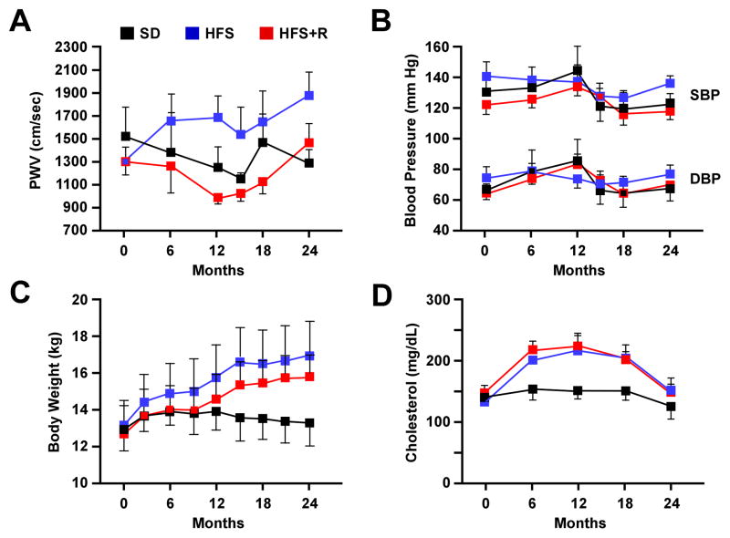
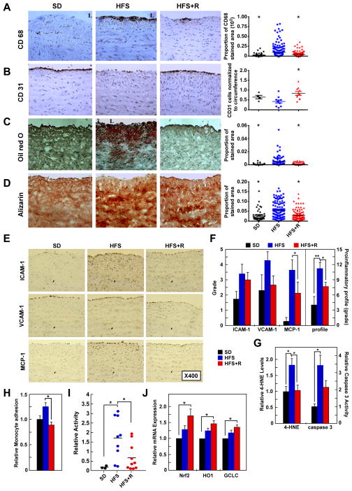
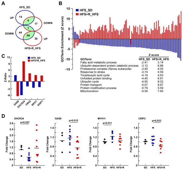
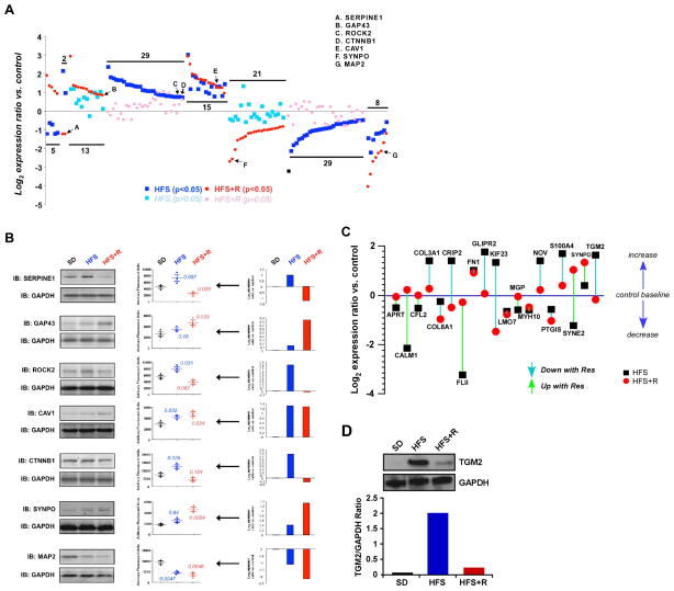
Central arterial wall stiffening, driven by a chronic inflammatory milieu, accompanies arterial diseases, the leading cause of cardiovascular (CV) morbidity and mortality in Western society. An increase in central arterial wall stiffening, measured as an increase in aortic pulse wave velocity (PWV), is a major risk factor for clinical CV disease events. However, no specific therapies to reduce PWV are presently available. In rhesus monkeys, a 2 year diet high in fat and sucrose (HFS) increases not only body weight and cholesterol, but also induces prominent central arterial wall stiffening and increases PWV and inflammation. The observed loss of endothelial cell integrity, lipid and macrophage infiltration, and calcification of the arterial wall were driven by genomic and proteomic signatures of oxidative stress and inflammation. Resveratrol prevented the HFS-induced arterial wall inflammation and the accompanying increase in PWV. Dietary resveratrol may hold promise as a therapy to ameliorate increases in PWV.


Conflict of interest statement

No potential conflicts of interest relevant to this article were reported.

Figures

Figure 1
Figure 1. Physiological Measurements in Rhesus Monkeys Maintained on HFS, HFS + R, or SD Diet for up to 2 Years
(A) Pulse wave velocity, (B) Blood pressure, (C) Body weight and (D) serum cholesterol levels. Results are mean ± SEM
Figure 2
Figure 2. Resveratrol Supplementation Retards the Adverse Molecular and Cellular Events within the Aortic Wall of Rhesus Monkeys on a HFS Diet and CAEC Incubation with Serum from HFS+Resv Serum Decreases Monocyte Adhesion
(A) Photomicrographs (X400) of the paraffin sections after immunostaining for CD68, a marker of monocyte/macrophage cells (left panel) (brown color determined by 3,3′-diaminobenzidine (DAB)); the average density of CD68-stained area/field/cross-section (right panel). (B) Photomicrographs (X400) of paraffin sections after immunostaining for CD31, a marker of endothelial cells (left panel); the number of CD31-stained endothelial cells normalized by the circumference (right panel). (C) Photomicrographs (X400) of frozen sections after Oil O Red staining (left panel); the density of lipid deposition/field/cross-section (right panel). (D) Photomicrographs (X400) of frozen sections after Alizarin red staining (left panel); the density of calcification/field/cross-section (right panel). For A–D, * p < 0.05 versus HFS. (E) Photomicrographs (X400) of frozen sections after immunostaining with antibodies for the proinflammatory molecules ICAM-1, VCAM-1, and MCP-1. (F) Grade average for ICAM-1, VCAM-1, and MCP-1 immunostaining as well as their summation (defined as local proinflammatory profile) of cells plus matrix within the intimal and medial compartment. (For AD, F: SD, n=4; HFS, n=10; HFS+R, n=10). (G) 4-HNE content and caspase 3 activity in aorta (SD, n=3; HFS, n=9; HFS+R, n=9). (H) Monocyte adhesion assay in human coronary artery endothelial cells (CAECs) cultured for 24 h with 10% serum from SD-, HFS- or HFS+R-fed rhesus monkeys. (I) NF-κB reporter assays were initiated after a 24-h incubation of human CAECs with HFS monkey serum. (J) Expression of Nrf2, HO1 and GCLC mRNA in human CAECs was determined by quantitative RT-PCR analysis. The effects of a 24-h incubation with HFS and HFS+R monkey serum were normalized to SD serum (for H–J: SD, n=3; HFS, n=9; HFS+R, n=10), for F–J: *, ** p< 0.05 and <0.01, respectively. Data are mean ± SEM for all graphs.
Figure 3
Figure 3. Resveratrol Supplementation Reverses Global Transcriptional Effects of a HFS in Aorta of Rhesus Monkeys
(A) Venn diagram of GO terms significantly affected by HFS diet vs. SD, and HFS+R vs. HFS. (B) Graphical representation of the significant 106 GO terms shared by HFS– (plotted in blue) and HFS+R –fed animals (plotted in red). Metabolic and catabolic processes, stress response and mitochondrion were among the regulated GO terms in aorta. Full GO term listing is provided in the Supplementary material. (C) Change in expression of select genes between HFS vs. SD and HFS+R vs. HFS is depicted as Z-ratios. List of genes whose expression in the HFS-fed group was reversed significantly by Resv supplementation is provided in the Supplemental material. (D) Validation of the microarray data by quantitative RT-PCR.
Figure 4
Figure 4. Resveratrol Supplementation Restores the Conversion of ‘Multidimensional’ Protein Network of the Cytoskeletal Dynamics and Vascular Functionality
(A) Log2-transformed SILAC expression ratio data for proteins extracted and identified in primary cultures of vascular smooth muscle (VSM) from rhesus monkey on SD, HFS and HFS+R diet. These proteins were selected due to their implicit textual association with vascular functionality (see Supplemental material for additional information). The proteins identified demonstrated a significantly altered degree of expression regulation in either the HFS (blue squares) or HFS+R (red circle) cohort (p<0.05). Non-significant expression changes are identified for the HFS cohort with light blue squares and for the HFS+R cohort with pink circles. (B) Western blot validation of several proteins identified with SILAC. Representative Western blot panels display the expression profile from a set of SD, HFS and HFS+R-fed monkeys. The associated scatter-plot histograms indicate the degree of individual animal variation in expression, as well as the significant p value (t-test) for HFS or HFS+R compared to SD. (C) The effects of Resv and HFS diet treatments upon nineteen ‘multidimensional’ proteins identified from multiple bioinformatic interpretation (GO-bp, KEGG, WikiPathways, PathwayCommons). These proteins isolated from VSM extracts were associated with all the interrogator terms. (D) TGM2 expression in primary culture of rhesus monkey VSM cells was measured using Western blot and expression levels were normalized to GAPDH.

References

    1. Bakker EN, Pistea A, VanBavel E. Transglutaminases in vascular biology: relevance for vascular remodeling and atherosclerosis. J Vasc Res. 2008;45:271–278. - PubMed
    1. Carrizzo A, Puca A, Damato A, Marino M, Franco E, Pompeo F, Traficante A, Civitillo F, Santini L, Trimarco V, et al. Resveratrol improves vascular function in patients with hypertension and dyslipidemia by modulating NO metabolism. Hypertension. 2013;62:359–366. - PubMed
    1. Chadwick W, Keselman A, Park SS, Zhou Y, Wang L, Brenneman R, Martin B, Maudsley S. Repetitive peroxide exposure reveals pleiotropic mitogen-activated protein kinase signaling mechanisms. J Signal Transduct. 2011;2011:636951. - PMC - PubMed
    1. Csiszar A, Sosnowska D, Wang M, Lakatta EG, Sonntag WE, Ungvari Z. Age-associated proinflammatory secretory phenotype in vascular smooth muscle cells from the non-human primate Macaca mulatta: reversal by resveratrol treatment. J Gerontol A Biol Sci Med Sci. 2012;67:811–820. - PMC - PubMed
    1. Do GM, Kwon EY, Kim HJ, Jeon SM, Ha TY, Park T, Choi MS. Long-term effects of resveratrol supplementation on suppression of atherogenic lesion formation and cholesterol synthesis in apo E-deficient mice. Biochem Biophys Res Commun. 2008;374:55–59. - PubMed

Publication types

MeSH terms