

Amygdala-prefrontal cortex functional connectivity during threat-induced anxiety and goal distraction
- PMID: 24882566
- PMCID: PMC4349396
- DOI: 10.1016/j.biopsych.2014.03.030
Amygdala-prefrontal cortex functional connectivity during threat-induced anxiety and goal distraction
Abstract
Background: Anxiety produced by environmental threats can impair goal-directed processing and is associated with a range of psychiatric disorders, particularly when aversive events occur unpredictably. The prefrontal cortex (PFC) is thought to implement controls that minimize performance disruptions from threat-induced anxiety and goal distraction by modulating activity in regions involved in threat detection, such as the amygdala. The inferior frontal gyrus (IFG), orbitofrontal cortex (OFC), and ventromedial PFC (vmPFC) have been linked to the regulation of anxiety during threat exposure. We developed a paradigm to determine if threat-induced anxiety would enhance functional connectivity between the amygdala and IFG, OFC, and vmPFC.
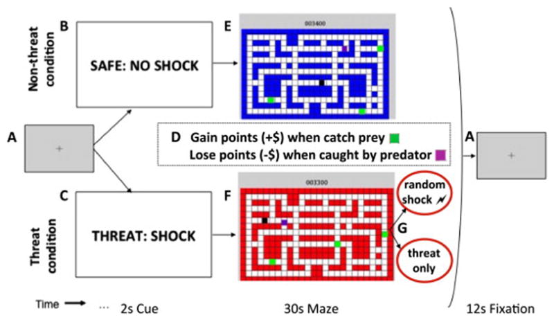
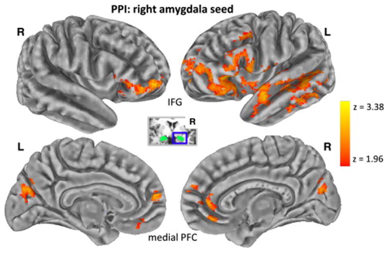
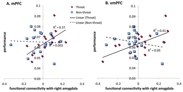
Methods: Healthy adults performed a computer-gaming style task involving capturing prey and evading predators to optimize monetary rewards while exposed to the threat of unpredictable shock. Psychophysiological recording (n = 26) and functional magnetic resonance imaging scanning (n = 17) were collected during the task in separate cohorts. Task-specific changes in functional connectivity with the amygdala were examined using psychophysiological interaction analysis.
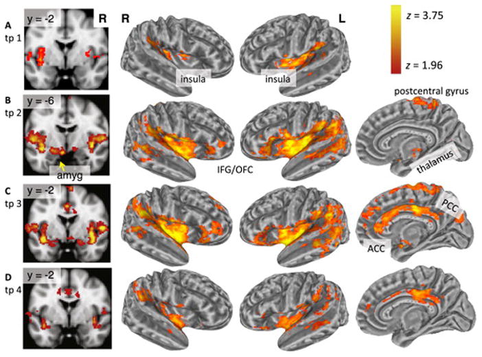
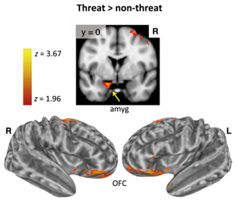
Results: Threat exposure resulted in greater arousal measured by increased skin conductance but did not influence performance (i.e., monetary losses or rewards). Greater functional connectivity between the right amygdala and bilateral IFG, OFC, vmPFC, anterior cingulate cortex, and frontopolar cortex was associated with threat exposure.
Conclusions: Exposure to unpredictable threat modulates amygdala-PFC functional connectivity that may help maintain performance when experiencing anxiety induced by threat. Our paradigm is well-suited to explore the neural underpinnings of the anxiety response to unpredictable threat in patients with various anxiety disorders.
Keywords: Amygdala; Functional connectivity; Inferior frontal gyrus; Orbitofrontal cortex; Psychophysiological interaction; Ventromedial prefrontal cortex.
Copyright © 2015. Published by Elsevier Inc.
Conflict of interest statement
Drs. Gold, Morey, and McCarthy have no biomedical financial interests or potential conflicts of interest to declare related to the present study.
Figures
Similar articles
-
Threat-induced anxiety during goal pursuit disrupts amygdala-prefrontal cortex connectivity in posttraumatic stress disorder.Transl Psychiatry. 2020 Feb 10;10(1):61. doi: 10.1038/s41398-020-0739-4. Transl Psychiatry. 2020. PMID: 32066690 Free PMC article.
-
Clinically Anxious Individuals Show Disrupted Feedback between Inferior Frontal Gyrus and Prefrontal-Limbic Control Circuit.J Neurosci. 2016 Apr 27;36(17):4708-18. doi: 10.1523/JNEUROSCI.1092-15.2016. J Neurosci. 2016. PMID: 27122030 Free PMC article.
-
Amygdala-Cortical Connectivity: Associations with Anxiety, Development, and Threat.Depress Anxiety. 2016 Oct;33(10):917-926. doi: 10.1002/da.22470. Depress Anxiety. 2016. PMID: 27699940 Free PMC article.
-
Emotion, motivation, decision-making, the orbitofrontal cortex, anterior cingulate cortex, and the amygdala.Brain Struct Funct. 2023 Jun;228(5):1201-1257. doi: 10.1007/s00429-023-02644-9. Epub 2023 May 13. Brain Struct Funct. 2023. PMID: 37178232 Free PMC article. Review.
-
The prefrontal cortex, pathological anxiety, and anxiety disorders.Neuropsychopharmacology. 2022 Jan;47(1):260-275. doi: 10.1038/s41386-021-01109-z. Epub 2021 Aug 16. Neuropsychopharmacology. 2022. PMID: 34400783 Free PMC article. Review.
Cited by
-
Neural and behavioral alterations of a real-time interpersonal distance (IPD) development process in differing social status interactions.Front Behav Neurosci. 2022 Oct 13;16:969440. doi: 10.3389/fnbeh.2022.969440. eCollection 2022. Front Behav Neurosci. 2022. PMID: 36311868 Free PMC article.
-
Predicting Brain Functional Connectivity Using Mobile Sensing.Proc ACM Interact Mob Wearable Ubiquitous Technol. 2020 Mar;4(1):23. doi: 10.1145/3381001. Epub 2020 Mar 18. Proc ACM Interact Mob Wearable Ubiquitous Technol. 2020. PMID: 36540188 Free PMC article.
-
The relationship between dlPFC activity during unpredictable threat and CO2-induced panic symptoms.Transl Psychiatry. 2017 Nov 30;7(12):1266. doi: 10.1038/s41398-017-0006-5. Transl Psychiatry. 2017. PMID: 29213110 Free PMC article.
-
Brain mechanisms of anxiety's effects on cognitive control in major depressive disorder.Psychol Med. 2016 Aug;46(11):2397-409. doi: 10.1017/S0033291716001185. Epub 2016 Jun 13. Psychol Med. 2016. PMID: 27291341 Free PMC article.
-
Antagonism between brain regions relevant for cognitive control and emotional memory facilitates the generation of humorous ideas.Sci Rep. 2021 May 21;11(1):10685. doi: 10.1038/s41598-021-89843-8. Sci Rep. 2021. PMID: 34021200 Free PMC article.
References
-
- Arnsten AF, Goldman-Rakic PS. Noise stress impairs prefrontal cortical cognitive function in monkeys: Evidence for a hyperdopaminergic mechanism. Arch Gen Psychiatry. 1998;55:362–368. - PubMed
-
- Eysenck MW, Derakshan N, Santos R, Calvo MG. Anxiety and cognitive performance: Attentional control theory. Emotion. 2007;7:336–353. - PubMed
-
- Bishop S, Duncan J, Brett M, Lawrence AD. Prefrontal cortical function and anxiety: Controlling attention to threat-related stimuli. Nat Neurosci. 2004;7:184–188. - PubMed
Publication types
MeSH terms
Grants and funding
LinkOut - more resources
Full Text Sources
Other Literature Sources
Medical
Miscellaneous
