. 2014 Oct:114:101-112.
doi: 10.1016/j.nlm.2014.05.005. Epub 2014 May 29.

A presynaptic role for PKA in synaptic tagging and memory

Affiliations

A presynaptic role for PKA in synaptic tagging and memory

Alan Jung Park et al. Neurobiol Learn Mem. 2014 Oct.

Abstract

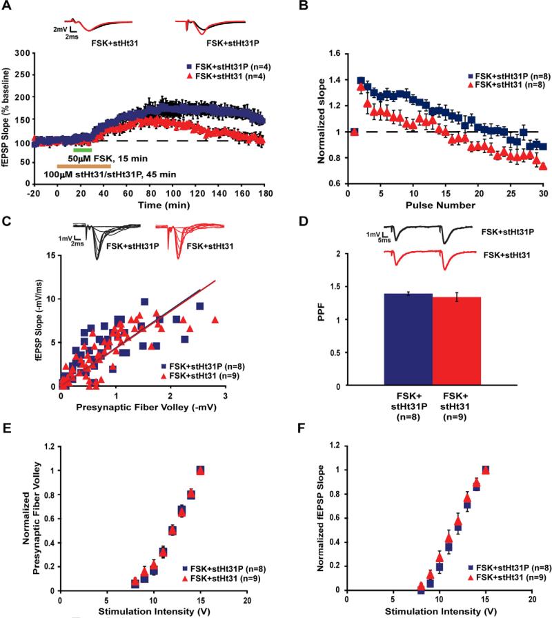
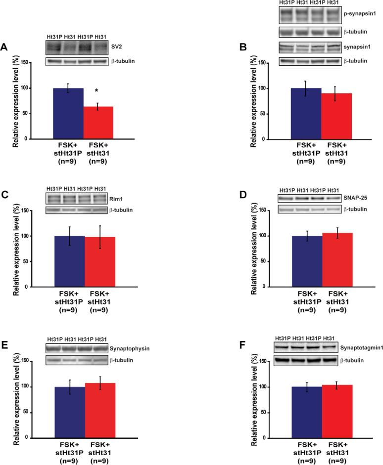
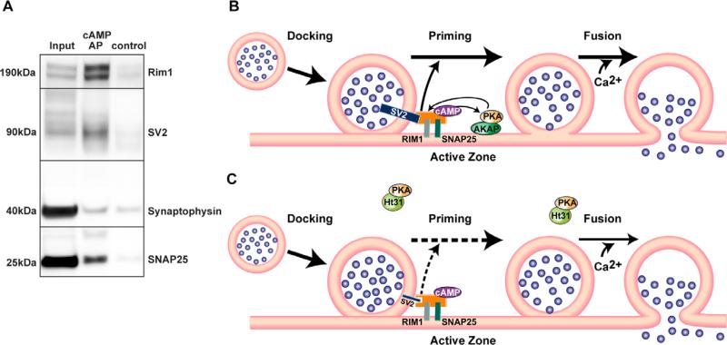
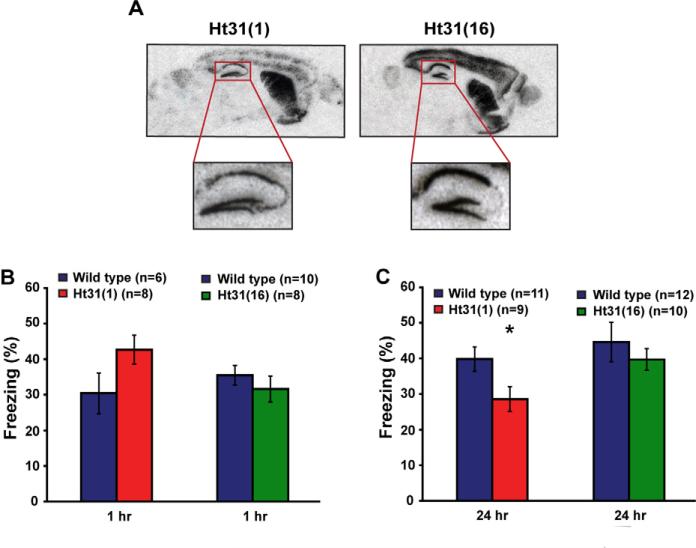
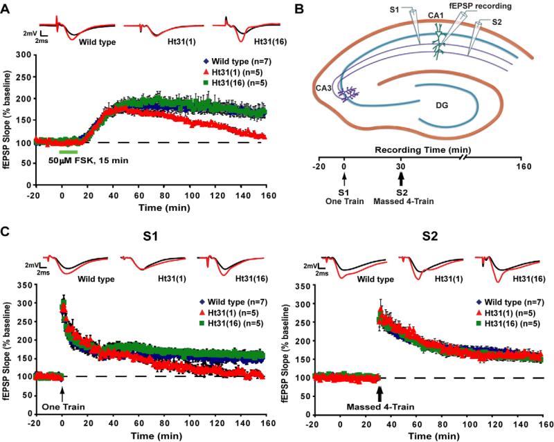
Protein kinase A (PKA) and other signaling molecules are spatially restricted within neurons by A-kinase anchoring proteins (AKAPs). Although studies on compartmentalized PKA signaling have focused on postsynaptic mechanisms, presynaptically anchored PKA may contribute to synaptic plasticity and memory because PKA also regulates presynaptic transmitter release. Here, we examine this issue using genetic and pharmacological application of Ht31, a PKA anchoring disrupting peptide. At the hippocampal Schaffer collateral CA3-CA1 synapse, Ht31 treatment elicits a rapid decay of synaptic responses to repetitive stimuli, indicating a fast depletion of the readily releasable pool of synaptic vesicles. The interaction between PKA and proteins involved in producing this pool of synaptic vesicles is supported by biochemical assays showing that synaptic vesicle protein 2 (SV2), Rim1, and SNAP25 are components of a complex that interacts with cAMP. Moreover, acute treatment with Ht31 reduces the levels of SV2. Finally, experiments with transgenic mouse lines, which express Ht31 in excitatory neurons at the Schaffer collateral CA3-CA1 synapse, highlight a requirement for presynaptically anchored PKA in pathway-specific synaptic tagging and long-term contextual fear memory. These results suggest that a presynaptically compartmentalized PKA is critical for synaptic plasticity and memory by regulating the readily releasable pool of synaptic vesicles.

Keywords: Ht31; PKA; PKA anchoring; SV2; Synaptic tagging.


Figures

Figure 1
Figure 1
Pharmacological disruption of PKA anchoring impairs forskolin (FSK)-induced long-lasting potentiation and facilitates short-term depression in the hippocampal Schaffer collateral pathway. A. Compared to the control stearated-Ht31P (stHt31P) peptide treatment (100 μM), stearated-Ht31 (stHt31) peptide treatment (100 μM) impairs a form of long-lasting potentiation induced by 15 minutes of bath application of 50 μM FSK (two-way repeated-measures ANOVA, F(1,6) = 15.045, p = 0.008). Representative traces acquired at the baseline (black) and at the last 20 minutes of the recordings (red) are shown on top of the graph. B. In FSK-treated slices, application of stHt31 peptide facilitated short-term depression induced by 10 Hz stimulation relative to application of stHt31P peptide (two-way repeated-measures ANOVA, F(1,14) = 8.377, p = 0.012). Values were normalized to the initial response. C. Slices co-treated with FSK and stHt31 peptide showed input-output relationships similar to slices co-treated with FSK and stHt31P peptide (t-test, p = 0.902). D. Paired-pulse facilitation (PPF) at 50 ms interval was not significantly different between FSK-treated slices perfused with stHt31 or stHt31P peptide (t-test, p = 0.46). E and F. Slices treated with stHt31 or stHt31P peptide along with FSK showed comparable changes in the presynaptic fiber volley (PFV) amplitude and initial fEPSP slope with increasing stimulation intensities (two-way repeated-measures ANOVA, for PFV amplitude: F(1,15) = 1.575, p = 0.606; for initial fEPSP slope: F(1,15) = 1.118, p = 0.295). Values were normalized to the maximum response. Error bars reflect S.E.M.
Figure 2
Figure 2
Pharmacological disruption of PKA anchoring selectively reduces the levels of synaptic vesicle protein 2 (SV2) in forskolin-treated hippocampal slices. A. stHt31 peptide treatment (100 μM) reduced the protein levels of SV2 relative to stHt31P peptide treatment (100 μM, t-test, p = 0.003). B. The ratio of phospho-synapsin1 to total synapsin1 was similar between slices treated with stHt31 or stHt31P (t-test, p = 0.6). The protein levels of other presynaptic proteins were not altered in slices treated with stHt31 peptide compared to slices treated with stHt31P peptide (C; Rim1: t-test, p = 0.94, D; SNAP25: t-test, p = 0.71, E; synaptophysin: t-test, p = 0.66, F; synaptotagmin1: t-test, p = 0.76). β-tubulin was used as the loading control and the expression level was normalized to the level of the control stHt31P peptide treatment group. Representative blots are shown on the left of each graph. * indicates p = 0.003. Error bars reflect S.E.M.
Figure 3
Figure 3
SV2, Rim1, and SNAP25 co-affinity precipitate with cAMP, and presynaptically compartmentalized PKA may regulate the readily releasable pool of synaptic vesicles through SV2. A. cAMP affinity-precipitation (AP) was performed using Rp-8-AHA-cAMP-agarose beads. Under control conditions, excessive amount of cAMP was added to compete against Rp-8-AHA-cAMP-agarose beads. cAMP affinity-precipitates of hippocampal extracts showed enrichment of Rim1, SV2, and SNAP25. The synaptophysin band in the AP lane was non-specific. B. A-kinase anchoring proteins (AKAPs) maintain a pool of PKA in close proximity to a cAMP-bound protein complex that bridges between a synaptic vesicle protein SV2 and active zone proteins, including Rim1 and SNAP25. This spatial compartmentalization allows fast coupling of cAMP-PKA signaling with the priming of the docked vesicles, which increases the size of the readily releasable pool of synaptic vesicles. Ca2+-influx following neuronal activation triggers fast fusion of the primed synaptic vesicles, and the large size of the readily releasable pool of synaptic vesicles sustains long-lasting synaptic plasticity. C. When PKA anchoring is disrupted by Ht31, PKA is sequestered away from the active zone. The free PKA is unable to coordinate interactions among proteins involved in vesicle exocytosis, which results in the reduction of SV2 protein levels. In addition, AKAPs may serve as the cAMP-bound complex, and Ht31 treatment disrupts the complex, thereby reducing SV2 levels. The reduced protein levels of SV2 destabilize the link between the docked synaptic vesicles and the active zone, thus the priming of the docked vesicles is attenuated. The resulting smaller size of the readily releasable pool of synaptic vesicles causes faster depletion of the vesicle pool during neuronal activity. Therefore, long-lasting synaptic plasticity is prevented. Note that this model focuses on how presynaptically compartmentalized PKA regulates the size of the readily releasable pool and does not exclude the potential involvement of other presynaptic proteins such as synapsin1 in the priming process.
Figure 4
Figure 4
Disruption of PKA anchoring in both CA3 and CA1 neurons, but not in CA1 neurons alone, impairs long-term contextual fear memory. A. Representative in situ hybridization images from sagittal brain sections demonstrated that both transgenic mouse lines expressed Ht31 in the major forebrain regions (upper panels). In the hippocampus, Ht31(1) mice expressed Ht31 throughout hippocampal subregions including the dentate gyrus, CA3, and CA1. Ht31(16) mice expressed Ht31 sparsely in CA3, but robustly in the dentate gyrus and CA1 (lower panels). B. Ht31(1) mice and wild-type littermates showed similar freezing levels during the retention test 1 hour after training (t-test, p = 0.076). Compared to wild-type littermates, Ht31(16) mice exhibited comparable freezing levels during the retention test 1 hour after training (t-test, p = 0.37). C. Ht31(1) mice displayed significantly decreased freezing levels during the retention test 24 hours after training relative to wild-type littermates (t-test, p = 0.033). However, Ht31(16) mice and wild-type littermates exhibited similar freezing levels during the retention test 24 hours after training (t-test, p = 0.452). * indicates p = 0.033. Error bars reflect S.E.M.
Figure 5
Figure 5
Disruption of PKA anchoring in both presynaptic CA3 and postsynaptic CA1 neurons, but not in postsynaptic CA1 neurons alone, impairs forskolin (FSK)-induced long-lasting potentiation and synaptic tagging. A. Ht31(16) mice and wild-type littermates, but not Ht31(1) mice, showed a form of long-lasting potentiation induced by 15 minutes of bath application of 50 μM FSK at the hippocampal Schaffer collateral CA3-CA1 synapses (two-way repeated-measures ANOVA, F(2,14) = 11.373, p = 0.001, Dunnett's post hoc test, Ht31(1) vs. wild-type p = 0.001, Ht31(16) vs. wild-type p = 0.99). B. A schematic diagram of the two-pathway experiment. Two stimulating electrodes were placed on either side of the recording electrode at the Schaffer collateral CA3-CA1 synapses to activate two independent sets of inputs (S1 and S2) onto the same postsynaptic population of neurons in CA1. Weak one train stimulation (one 1s 100 Hz train) was delivered to S1 first, and 30 minutes later, strong massed 4-train stimulation (four 1s 100 Hz trains with 5 seconds intertrain interval) was delivered to S2. C. Synaptic tagging is impaired in hippocampal slices from Ht31(1), but not in Ht31(16) mice. In S1, weak one train stimulation induced long-lasting potentiation in hippocampal slices from both wild-type littermates and Ht31(16) mice, but not in slices from Ht31(1) mice (Left panel; two-way repeated-measures ANOVA, F(2,14) = 26.139, p = 0.00002, Dunnett's post hoc test, Ht31(1) vs. wild-type p = 0.00011, Ht31(16) vs. wild-type p = 0.195). In S2, strong massed 4-train stimulation elicited similar levels of long-lasting LTP in slices from Ht31(1), Ht31(16) mice, and wild-type littermates (Right panel; two-way repeated-measures ANOVA, F(2,14) = 0.2, p = 0.821). Representative traces acquired at the baseline (black) and at the last 20 minutes of the recordings (red) are shown on top of the graph. Error bars reflect S.E.M.

Similar articles

Cited by

References

    1. Ahmari SE, Buchanan J, Smith SJ. Assembly of presynaptic active zones from cytoplasmic transport packets. Nature Neuroscience. 2000;3(5):445–51. - PubMed
    1. Baba T, Sakisaka T, Mochida S, Takai Y. PKA-catalyzed phosphorylation of tomosyn and its implication in Ca2+-dependent exocytosis of neurotransmitter. The Journal of Cell Biology. 2005;170(7):1113–25. - PMC - PubMed
    1. Bajjalieh SM, Frantz GD, Weimann JM, McConnell SK, Scheller RH. Differential expression of synaptic vesicle protein 2 (SV2) isoforms. The Journal of Neuroscience: The Official Journal of the Society for Neuroscience. 1994;14(9):5223–35. - PMC - PubMed
    1. Bajjalieh SM, Peterson K, Linial M, Scheller RH. Brain contains two forms of synaptic vesicle protein 2. Proceedings of the National Academy of Sciences of the United States of America. 1993;90(6):2150–4. - PMC - PubMed
    1. Barco A, Alarcon JM, Kandel ER. Expression of constitutively active CREB protein facilitates the late phase of long-term potentiation by enhancing synaptic capture. Cell. 2002;108(5):689–703. - PubMed

Publication types

MeSH terms