

Exosomes mediate intercellular transfer of pro-fibrogenic connective tissue growth factor (CCN2) between hepatic stellate cells, the principal fibrotic cells in the liver
- PMID: 24882759
- PMCID: PMC4150742
- DOI: 10.1016/j.surg.2014.04.014
Exosomes mediate intercellular transfer of pro-fibrogenic connective tissue growth factor (CCN2) between hepatic stellate cells, the principal fibrotic cells in the liver
Abstract
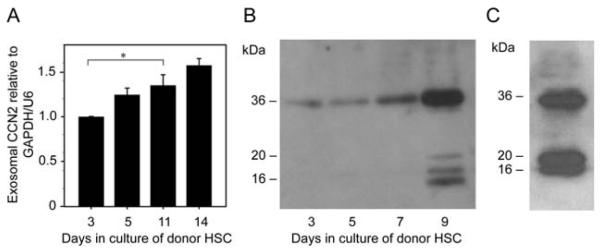
Background: Fibrogenic pathways in the liver are principally regulated by hepatic stellate cells (HSC), which produce and respond to fibrotic mediators such as connective tissue growth factor (CCN2). The aim of this study was to determine whether CCN2 is shuttled between HSC in membranous nanovesicles, or "exosomes."
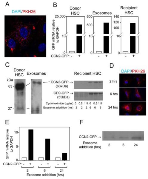
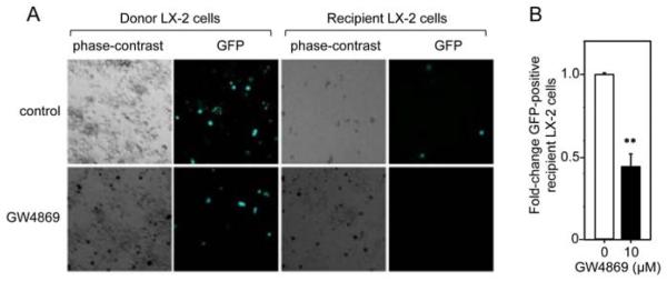
Methods: Exosomes were incubated with HSC after isolation from conditioned medium of control or CCN2-green fluorescent protein (GFP)-transfected primary mouse HSC or human LX-2 HSC. Some exosomes were stained fluorescently with PKH26. HSC co-culture experiments were performed in the presence of GW4869 exosome inhibitor. CCN2 or CCN2-GFP were evaluated by quantitative real-time polymerase chain reaction or Western blot.
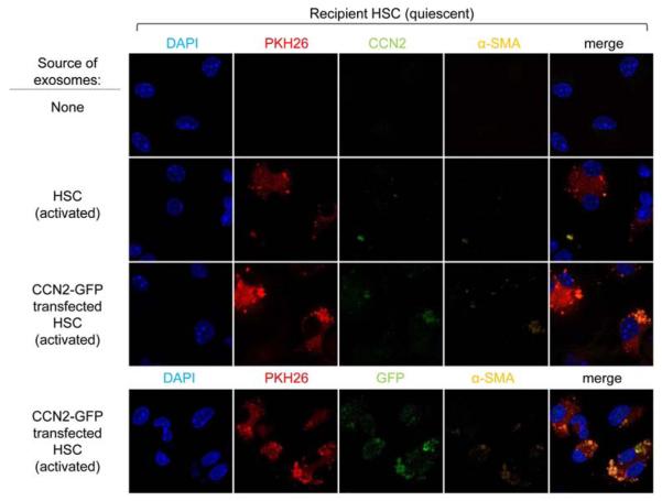
Results: HSC-derived exosomes contained CCN2 or CCN2 mRNA, each of which increased in concentration during HSC activation or after transfection of HSC with CCN2-GFP. Exosomes, stained with either PKH26 or purified from CCN2-GFP-transfected cells, were taken up by activated or quiescent HSC resulting in CCN2-GFP delivery, as shown by their direct addition to recipient cells or by the GW4869-dependency of donor HSC.
Conclusion: CCN2 is packaged into secreted, nano-sized exosomes that mediate its intercellular transfer between HSC. Exosomal CCN2 may amplify or fine tune fibrogenic signaling and, in conjunction with other exosome constituents, may have utility as a noninvasive biomarker to assess hepatic fibrosis.
Copyright © 2014 Mosby, Inc. All rights reserved.
Figures
References
-
- Eng FJ, Friedman SL, Fibrogenesis I. New insights into hepatic stellate cell activation: the simple becomes complex. Am J Physiol Gastrointest Liver Physiol. 2000;279:G7–G11. - PubMed
-
- Friedman SL. Molecular regulation of hepatic fibrosis, an integrated cellular response to tissue injury. J Biol Chem. 2000;275:2247–50. - PubMed
-
- Gaca MD, Zhou X, Benyon RC. Regulation of hepatic stellate cell proliferation and collagen synthesis by proteinase-activated receptors. J Hepatol. 2002;36:362–9. - PubMed
-
- Friedman SL. Hepatic fibrosis -- overview. Toxicology. 2008;254:120–9. - PubMed
Publication types
MeSH terms
Substances
Grants and funding
LinkOut - more resources
Full Text Sources
Other Literature Sources
Miscellaneous
