

Bronchoalveolar lavage fluid IFN-γ+ Th17 cells and regulatory T cells in pulmonary sarcoidosis
- PMID: 24882950
- PMCID: PMC4027000
- DOI: 10.1155/2014/438070
Bronchoalveolar lavage fluid IFN-γ+ Th17 cells and regulatory T cells in pulmonary sarcoidosis
Abstract
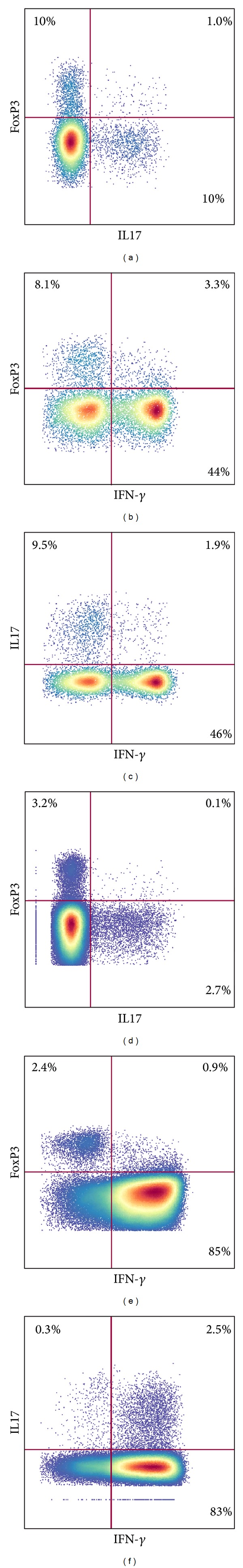
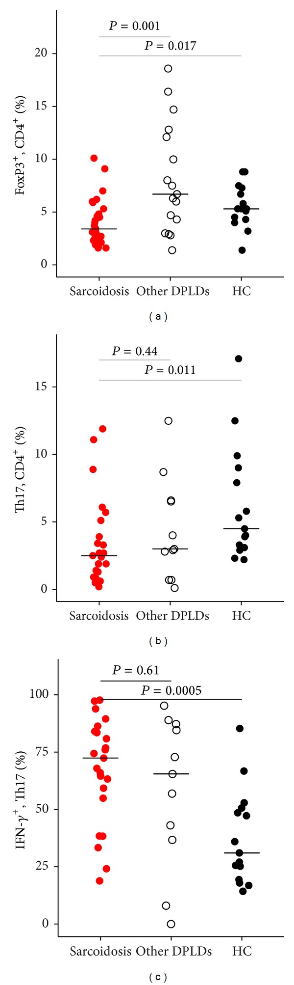
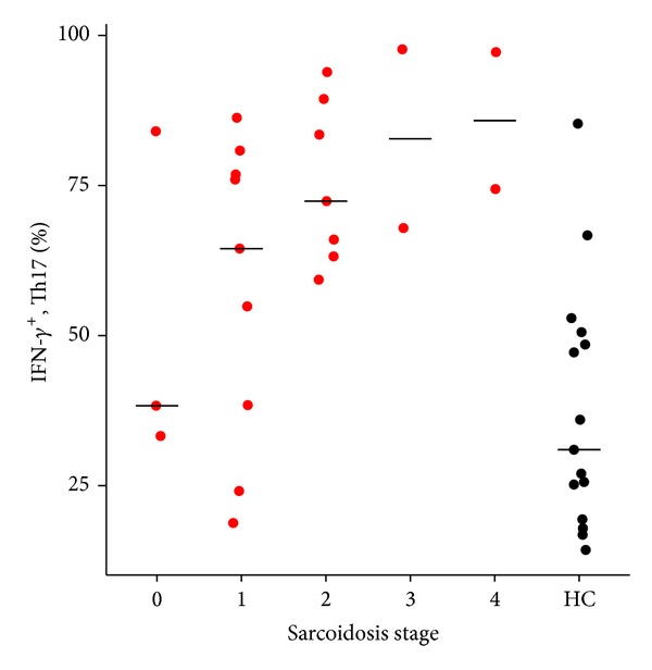
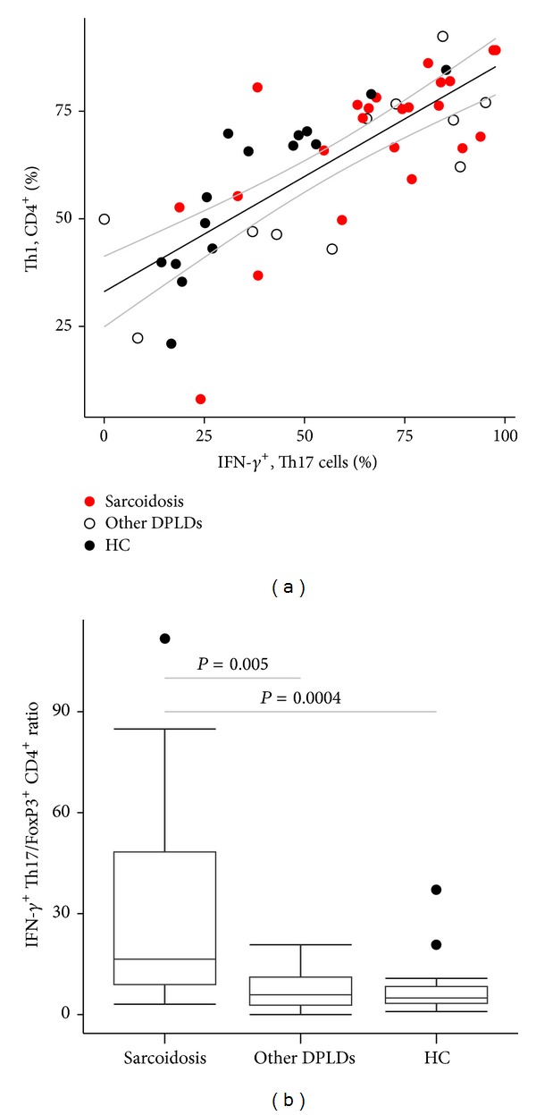
In sarcoidosis, increased Th17 cell fractions have been reported in bronchoalveolar lavage fluid, and elevated numbers of Th17 cells producing IFN- γ have been observed in peripheral blood. The balance between Th1, Th17, and FoxP3(+) CD4(+) T cell subsets in sarcoidosis remains unclear. Bronchoalveolar lavage fluid cells, from 30 patients with sarcoidosis, 18 patients with other diffuse parenchymal lung diseases, and 15 healthy controls, were investigated with flow cytometry for intracellular expression of FoxP3. In a subset of the patients, expression of the cytokines IL17A and IFN- γ was investigated. The fractions of FoxP3(+) CD4(+) T cells and Th17 cells were both lower in sarcoidosis compared to controls (P = 0.017 and P = 0.011, resp.). The proportion of Th17 cells positive for IFN- γ was greater in sarcoidosis than controls (median 72.4% versus 31%, P = 0.0005) and increased with radiologic stage (N = 23, rho = 0.45, and P = 0.03). IFN- γ (+) Th17 cells were highly correlated with Th1 cells (N = 23, rho = 0.64, and P = 0.001), and the ratio of IFN- γ (+) Th17/FoxP3(+) CD4(+) T cells was prominently increased in sarcoidosis. IFN- γ (+) Th17 cells may represent a pathogenic subset of Th17 cells, yet their expression of IFN- γ could be a consequence of a Th1-polarized cytokine milieu. Our results indicate a possible immune cell imbalance in sarcoidosis.
Figures
Similar articles
-
IFN-γ-Producing T-Helper 17.1 Cells Are Increased in Sarcoidosis and Are More Prevalent than T-Helper Type 1 Cells.Am J Respir Crit Care Med. 2016 Jun 1;193(11):1281-91. doi: 10.1164/rccm.201507-1499OC. Am J Respir Crit Care Med. 2016. PMID: 26649486 Free PMC article.
-
Expanded lung T-bet+RORγT+ CD4+ T-cells in sarcoidosis patients with a favourable disease phenotype.Eur Respir J. 2016 Aug;48(2):484-94. doi: 10.1183/13993003.00092-2016. Epub 2016 May 26. Eur Respir J. 2016. PMID: 27230441
-
T-bet Expression in Peripheral Th17.0 Cells Is Associated With Pulmonary Function Changes in Sarcoidosis.Front Immunol. 2020 Jul 22;11:1129. doi: 10.3389/fimmu.2020.01129. eCollection 2020. Front Immunol. 2020. PMID: 32774332 Free PMC article.
-
The Roles of T Helper 1, T Helper 17 and Regulatory T Cells in the Pathogenesis of Sarcoidosis.Iran J Allergy Asthma Immunol. 2016 Aug;15(4):334-339. Iran J Allergy Asthma Immunol. 2016. PMID: 27921415 Review.
-
Th17-lineage cells in pulmonary sarcoidosis and Löfgren's syndrome: Friend or foe?J Autoimmun. 2018 Feb;87:82-96. doi: 10.1016/j.jaut.2017.12.012. Epub 2018 Jan 5. J Autoimmun. 2018. PMID: 29310925 Review.
Cited by
-
The Role of Diverse Immune Cells in Sarcoidosis.Front Immunol. 2021 Nov 19;12:788502. doi: 10.3389/fimmu.2021.788502. eCollection 2021. Front Immunol. 2021. PMID: 34868074 Free PMC article. Review.
-
A Gene-Environment Interaction Between Smoking and Gene polymorphisms Provides a High Risk of Two Subgroups of Sarcoidosis.Sci Rep. 2019 Dec 9;9(1):18633. doi: 10.1038/s41598-019-54612-1. Sci Rep. 2019. PMID: 31819081 Free PMC article.
-
Follicular T Cells from smB- Common Variable Immunodeficiency Patients Are Skewed Toward a Th1 Phenotype.Front Immunol. 2017 Feb 27;8:174. doi: 10.3389/fimmu.2017.00174. eCollection 2017. Front Immunol. 2017. PMID: 28289412 Free PMC article.
-
Reduced expression of peroxisome proliferator-activated receptor alpha in BAL and blood T cells of non-löfgren's sarcoidosis patients.J Inflamm (Lond). 2015 Apr 9;12:28. doi: 10.1186/s12950-015-0071-6. eCollection 2015. J Inflamm (Lond). 2015. PMID: 25969669 Free PMC article.
-
Circulating Regulatory T Cell Subsets in Patients with Sarcoidosis.Diagnostics (Basel). 2023 Apr 10;13(8):1378. doi: 10.3390/diagnostics13081378. Diagnostics (Basel). 2023. PMID: 37189479 Free PMC article.
References
-
- Chen ES, Moller DR. Sarcoidosis—scientific progress and clinical challenges. Nature Reviews Rheumatology. 2011;7(8):457–467. - PubMed
-
- Prince JE, Kheradmand F, Corry DB. 16. Immunologic lung disease. Journal of Allergy and Clinical Immunology. 2003;111(2, supplement 2):S613–S623. - PubMed
-
- Zissel G, Prasse A, Muller-Quernheim J. Immunologic response of sarcoidosis. Seminars in Respiratory and Critical Care Medicine. 2010;31(4):390–403. - PubMed
Publication types
MeSH terms
Substances
LinkOut - more resources
Full Text Sources
Other Literature Sources
Research Materials
