

Photodynamic Therapy for Bowen's Disease of the Vulva Area
- PMID: 24882981
- PMCID: PMC4037679
- DOI: 10.5021/ad.2014.26.2.241
Photodynamic Therapy for Bowen's Disease of the Vulva Area
Abstract
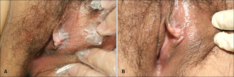
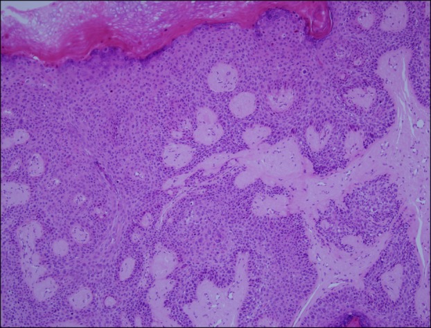
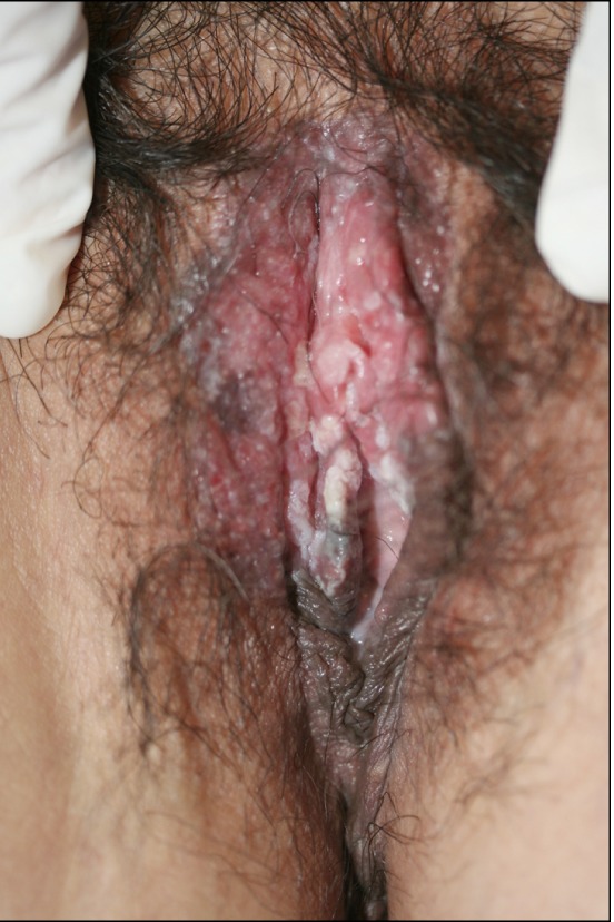
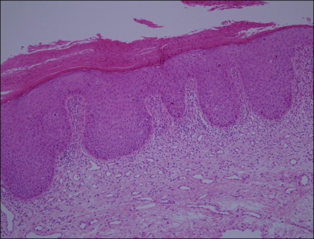
Bowen's disease is a squamous cell carcinoma in situ and has the potential to progress to a squamous cell carcinoma. The authors treated two female patients (a 39-year-old and a 41-year-old) with Bowen's disease in the vulva area using topical photodynamic therapy (PDT), involving the use of 5-aminolaevulinic acid and a light-emitting diode device. The light was administered at an intensity of 80 mW/cm(2) for a dose of 120 J/cm(2) biweekly for 6 cycles. The 39-year-old patient showed excellent clinical improvement, but the other patient achieved only a partial response. Even though one patient underwent a total excision 1 year later due to recurrence, both patients were satisfied with the cosmetic outcomes of this therapy and the partial improvement over time. The common side effect of PDT was a stinging sensation. PDT provides a relatively effective and useful alternative treatment for Bowen's disease in the vulva area.
Keywords: Bowen's disease; Photochemotherapy; Vulva area.
Figures
Similar articles
-
Bowen's disease of the vulva and perianal area in a SLE patient successfully treated with Photodynamic therapy: A case report.Photodiagnosis Photodyn Ther. 2024 Feb;45:103910. doi: 10.1016/j.pdpdt.2023.103910. Epub 2023 Nov 30. Photodiagnosis Photodyn Ther. 2024. PMID: 38042234
-
Photodynamic therapy with violet light and topical 6-aminolaevulinic acid in the treatment of actinic keratosis, Bowen's disease and basal cell carcinoma.J Eur Acad Dermatol Venereol. 2001 Nov;15(6):550-4. doi: 10.1046/j.1468-3083.2001.00333.x. J Eur Acad Dermatol Venereol. 2001. PMID: 11843215 Clinical Trial.
-
Investigation of the use of the pulsed dye laser in the treatment of Bowen's disease using 5-aminolaevulinic acid phototherapy.Br J Dermatol. 2005 Oct;153(4):780-4. doi: 10.1111/j.1365-2133.2005.06830.x. Br J Dermatol. 2005. PMID: 16181460
-
Photosensitizers and light sources for photodynamic therapy of the Bowen's disease.Arch Dermatol Res. 2011 Apr;303(3):145-51. doi: 10.1007/s00403-011-1122-3. Epub 2011 Feb 1. Arch Dermatol Res. 2011. PMID: 21286736 Review.
-
Photodynamic therapy for Bowen's Disease (squamous cell carcinoma in situ) current review and update.Photodiagnosis Photodyn Ther. 2018 Dec;24:109-114. doi: 10.1016/j.pdpdt.2018.09.009. Epub 2018 Sep 18. Photodiagnosis Photodyn Ther. 2018. PMID: 30240928 Review.
Cited by
-
Vulvar cancer: a review for dermatologists.Wien Med Wochenschr. 2015 Apr;165(7-8):164-77. doi: 10.1007/s10354-015-0354-9. Epub 2015 May 1. Wien Med Wochenschr. 2015. PMID: 25930015 Review.
-
A new optical intra-tissue fiber irradiation ALA-PDT in the treatment of acne vulgaris in rabbit model: improved safety and tolerability.An Bras Dermatol. 2017 May-Jun;92(3):350-355. doi: 10.1590/abd1806-4841.20175543. An Bras Dermatol. 2017. PMID: 29186247 Free PMC article.
-
Susceptibility and Resistance Mechanisms During Photodynamic Therapy of Melanoma.Front Oncol. 2020 May 12;10:597. doi: 10.3389/fonc.2020.00597. eCollection 2020. Front Oncol. 2020. PMID: 32528867 Free PMC article. Review.
-
Nanoparticle-Based Drug Delivery Systems for Photodynamic Therapy of Metastatic Melanoma: A Review.Int J Mol Sci. 2021 Nov 21;22(22):12549. doi: 10.3390/ijms222212549. Int J Mol Sci. 2021. PMID: 34830431 Free PMC article. Review.
References
-
- Strayer DS, Santa Cruz DJ. Carcinoma in situ of the skin: a review of histopathology. J Cutan Pathol. 1980;7:244–259. - PubMed
-
- Duncan KO, Geisse JK, Leffell DJ. Epithelial precancerous lesions. In: Wolff K, Austen KF, Goldsmith LA, Katz SI, Gilchrest BA, Paller AS, et al., editors. Fitzpatrick's dermatology in general medicine. 7th ed. New York: McGraw-Hill; 2007. pp. 1021–1023.
-
- Svanberg K, Andersson T, Killander D, Wang I, Stenram U, Andersson-Engels S, et al. Photodynamic therapy of nonmelanoma malignant tumours of the skin using topical deltaamino levulinic acid sensitization and laser irradiation. Br J Dermatol. 1994;130:743–751. - PubMed
-
- Fijan S, Hönigsmann H, Ortel B. Photodynamic therapy of epithelial skin tumours using delta-aminolaevulinic acid and desferrioxamine. Br J Dermatol. 1995;133:282–288. - PubMed
-
- Morton CA, Whitehurst C, Moore JV, MacKie RM. Comparison of red and green light in the treatment of Bowen's disease by photodynamic therapy. Br J Dermatol. 2000;143:767–772. - PubMed
Publication types
LinkOut - more resources
Full Text Sources
Other Literature Sources
