Simultaneous mapping of pan and sentinel lymph nodes for real-time image-guided surgery
- PMID: 24883119
- PMCID: PMC4038751
- DOI: 10.7150/thno.8721
Simultaneous mapping of pan and sentinel lymph nodes for real-time image-guided surgery
Abstract
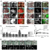
The resection of regional lymph nodes in the basin of a primary tumor is of paramount importance in surgical oncology. Although sentinel lymph node mapping is now the standard of care in breast cancer and melanoma, over 20% of patients require a completion lymphadenectomy. Yet, there is currently no technology available that can image all lymph nodes in the body in real time, or assess both the sentinel node and all nodes simultaneously. In this study, we report an optical fluorescence technology that is capable of simultaneous mapping of pan lymph nodes (PLNs) and sentinel lymph nodes (SLNs) in the same subject. We developed near-infrared fluorophores, which have fluorescence emission maxima either at 700 nm or at 800 nm. One was injected intravenously for identification of all regional lymph nodes in a basin, and the other was injected locally for identification of the SLN. Using the dual-channel FLARE intraoperative imaging system, we could identify and resect all PLNs and SLNs simultaneously. The technology we describe enables simultaneous, real-time visualization of both PLNs and SLNs in the same subject.
Keywords: Near-infrared fluorescence; cervical cancer.; completion lymphadenectomy; image-guided surgery; pan lymph node mapping; sentinel lymph node mapping.
Conflict of interest statement
Conflict of Interest: FLARE™ technology is owned by Beth Israel Deaconess Medical Center, a teaching hospital of Harvard Medical School. It has been licensed to the FLARE Foundation, a non-profit organization focused on promoting the dissemination of medical imaging technology for research and clinical use. Dr. Frangioni is the founder and chairman of the FLARE Foundation. The Beth Israel Deaconess Medical Center will receive royalties for sale of FLARE™ Technology. Dr. Frangioni has elected to surrender post-market royalties to which he would otherwise be entitled as inventor, and has elected to donate pre-market proceeds to the FLARE Foundation. Dr. Frangioni has started three for-profit companies, Curadel, Curadel ResVet Imaging, and Curadel Surgical Innovations, which may someday be non-exclusive sub-licensees of FLARE™ technology.
Figures


Similar articles
-
Intraoperative combined color and fluorescent images-based sentinel node mapping in the porcine lung: comparison of indocyanine green with or without albumin premixing.J Thorac Cardiovasc Surg. 2013 Dec;146(6):1509-15. doi: 10.1016/j.jtcvs.2013.02.044. Epub 2013 Mar 21. J Thorac Cardiovasc Surg. 2013. PMID: 23522603
-
Molecular Imaging of endometrial sentinel lymph nodes utilizing fluorescent-labeled Tilmanocept during robotic-assisted surgery in a porcine model.PLoS One. 2018 Jul 2;13(7):e0197842. doi: 10.1371/journal.pone.0197842. eCollection 2018. PLoS One. 2018. PMID: 29965996 Free PMC article.
-
Sentinel Node Mapping Using Indocyanine Green and Near-infrared Fluorescence Imaging Technology for Uterine Malignancies: Preliminary Experience With the Da Vinci Xi System.J Minim Invasive Gynecol. 2016 May-Jun;23(4):470-1. doi: 10.1016/j.jmig.2015.12.013. Epub 2016 Jan 6. J Minim Invasive Gynecol. 2016. PMID: 26767824
-
Near-infrared fluorescence imaging for sentinel lymph node identification in colon cancer: a prospective single-center study and systematic review with meta-analysis.Tech Coloproctol. 2019 Dec;23(12):1113-1126. doi: 10.1007/s10151-019-02107-6. Epub 2019 Nov 18. Tech Coloproctol. 2019. PMID: 31741099 Free PMC article.
-
Infrared Fluorescence-guided Surgery for Tumor and Metastatic Lymph Node Detection in Head and Neck Cancer.Radiol Imaging Cancer. 2024 Jul;6(4):e230178. doi: 10.1148/rycan.230178. Radiol Imaging Cancer. 2024. PMID: 38940689 Free PMC article. Review.
Cited by
-
Charge and hydrophobicity effects of NIR fluorophores on bone-specific imaging.Theranostics. 2015 Mar 1;5(6):609-17. doi: 10.7150/thno.11222. eCollection 2015. Theranostics. 2015. PMID: 25825600 Free PMC article.
-
Sentinel Lymph Node Mapping of Liver.Ann Surg Oncol. 2015 Dec;22 Suppl 3(0 3):S1147-55. doi: 10.1245/s10434-015-4601-5. Epub 2015 May 13. Ann Surg Oncol. 2015. PMID: 25968620 Free PMC article.
-
Phosphonated near-infrared fluorophores for biomedical imaging of bone.Angew Chem Int Ed Engl. 2014 Sep 26;53(40):10668-72. doi: 10.1002/anie.201404930. Epub 2014 Aug 19. Angew Chem Int Ed Engl. 2014. PMID: 25139079 Free PMC article.
-
Endocrine-specific NIR fluorophores for adrenal gland targeting.Chem Commun (Camb). 2016 Aug 11;52(67):10305-8. doi: 10.1039/c6cc03845j. Chem Commun (Camb). 2016. PMID: 27476533 Free PMC article.
-
Near-Infrared Illumination of Native Tissues for Image-Guided Surgery.J Med Chem. 2016 Jun 9;59(11):5311-23. doi: 10.1021/acs.jmedchem.6b00038. Epub 2016 May 19. J Med Chem. 2016. PMID: 27100476 Free PMC article.
References
-
- Morton DL, Wen DR, Wong JH, Economou JS, Cagle LA, Storm FK. et al. Technical details of intraoperative lymphatic mapping for early stage melanoma. Arch Surg. 1992;127:392–9. - PubMed
-
- Nieweg OE, van Rijk MC, Valdes Olmos RA, Hoefnagel CA. Sentinel node biopsy and selective lymph node clearance--impact on regional control and survival in breast cancer and melanoma. European journal of nuclear medicine and molecular imaging. 2005;32:631–4. - PubMed
-
- Chen SL, Iddings DM, Scheri RP, Bilchik AJ. Lymphatic mapping and sentinel node analysis: current concepts and applications. CA Cancer J Clin. 2006;56:292–309. quiz 16-7. - PubMed
-
- van der Pas MH, Meijer S, Hoekstra OS, Riphagen II, de Vet HC, Knol DL. et al. Sentinel-lymph-node procedure in colon and rectal cancer: a systematic review and meta-analysis. Lancet Oncol. 2011;12:540–50. - PubMed
-
- Lips DJ, Schutte HW, van der Linden RL, Dassen AE, Voogd AC, Bosscha K. Sentinel lymph node biopsy to direct treatment in gastric cancer. A systematic review of the literature. Eur J Surg Oncol. 2011;37:655–61. - PubMed
Publication types
MeSH terms
Substances
Grants and funding
LinkOut - more resources
Full Text Sources
Other Literature Sources

