. 2013 Aug 3;17(2):79-87.
doi: 10.1007/s40477-013-0025-x. eCollection 2014 Jun.

Sonographic anatomy of the ankle

Affiliations

Sonographic anatomy of the ankle

M Precerutti et al. J Ultrasound. .

Abstract

Ankle sonography is one of the most commonly ordered examinations in the field of osteoarticular imaging, and it requires intimate knowledge of the anatomic structures that make up the joint. For practical purposes, the examination can be divided into four compartments, which are analyzed in this pictorial essay: the anterior compartment, which includes the tibialis anterior, extensor hallucis longus, and extensor digitorum longus tendons; the accessory peroneus tertius tendon; and the extensor retinaculum; the medial compartment (tibialis posterior, flexor digitorum longus, and flexor hallucis longus tendons; the flexor retinaculum; the medial collateral-or deltoid-ligament, and the neurovascular bundle); the lateral compartment (peroneus longus, peroneus brevis, and peroneus quartus tendons; superior and inferior peroneal retinacula, lateral collateral ligament); and the posterior compartment (Achilles tendon, plantaris tendon, Kagar's triangle, superficial, and deep retrocalcaneal bursae). Scanning techniques are briefly described to ensure optimal visualization of the various anatomic structures.

L’esame ecografico della caviglia è tra gli esami più richiesti nell’ambito dell’ecografia osteoarticolare; ne deriva la necessità di una conoscenza approfondita delle strutture anatomiche che la compongono. L’approccio ecografico per lo studio della caviglia è, per scopi pratici, organizzato per comparti; si analizzano nel presente pictorial essay le strutture del comparto anteriore (tendine tibiale anteriore, tendine estensore lungo dell’alluce, tendine estensore lungo delle dita, tendine peroneo tertius, retinacolo degli estensori), del comparto mediale (tendine tibiale posteriore, tendine flessore lungo delle dita, tendine flessore lungo dell’alluce, retinacolo dei flessori, legamento collaterale mediale o deltoideo, fascio vascolo-nervoso), del comparto esterno (tendini peronei breve e lungo, tendine peroneo quarto, retinacolo superiore ed inferiore dei peronei, legamento collaterale esterno), del comparto posteriore (tendine d’Achille, tendine del muscolo plantare, triangolo di Kager, borse sinoviali calcaneali superficiale e profonda). Si riporta inoltre qualche breve cenno di tecnica ecografica necessaria per l’ottimale visualizzazione delle strutture descritte.

Keywords: Achilles tendon; Ankle; Extensor tendons of the foot; Flexor tendons of the foot; Ligaments of the ankle; Ultrasonography.


Figures

Fig. 1
Fig. 1
T1-weighted axial MR image showing the anterior compartment, which contains (lateral to medial): the tibialis anterior (arrowhead), the extensor hallucis longus (thick arrow), and the extensor digitorum comunis (thin arrow). The anteroinferior tibiofibular ligament can be seen near the tibiofibular syndesmosis (crooked arrow)
Fig. 2
Fig. 2
Anterior axial sonographic image (panoramic view) shows the three tendons of the anterior compartment (medial > lateral): the tibialis anterior (arrowhead), the extensor hallucis longus (thick arrow), and the extensor digitorum longus (thin arrow). The curved hyperechoic line deep to these structures represents the cortical bone of the anterior aspect of the distal tibial epiphysis
Fig. 3
Fig. 3
Anterior longitudinal sonographic image along the course of the tibialis anterior tendon (arrow) (panoramic view). The tendon itself, which has a typical fibrillar appearance, is seen running superficial to the anterior recess of the tibiotalar joint. The latter, which can be visualized only when it is distended secondary to effusion fluid, lies between the distal tibial epiphysis (curved arrow) and the talar dome (arrowhead)
Fig. 4
Fig. 4
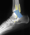
Anatomic diagram showing the superior and inferior extensor retinacula: the former stretches between the medial tibia and the distal fibula; the latter, which is Y-shaped, extends from the calcaneus to the medial malleolus and talus
Fig. 5
Fig. 5
Anterior axial sonographic image obtained at the level of the tibiofibular syndesmosis (panoramic image): the anteroinferior tibiofibular ligament is seen as a moderately thick echoic band on the anterior aspect of the joint (arrows)
Fig. 6
Fig. 6
T1-weighted axial MR image. a The medial compartment of the ankle contains the tibialis posterior (thick arrow), the flexor digitorum longus (thin arrow), the flexor hallucis longus (right-angled arrow) tendons; the tibial neurovascular bundle (curved arrow), which includes the tibial nerve, the tibial artery, and the posterior tibial veins. Coronal MR image (b) clearly depicts the superficial and deep fasciculi of the medial collateral (deltoid) ligament (straight arrow) and, superficially, the flexor retinaculum (curved arrow)
Fig. 7
Fig. 7
Sonographic image of the medial retromalleolar area obtained in the axial-oblique plane (panoramic image): a superficial to the bone plane, moving from the anterior to the posterior, one sees the tibialis posterior tendon (arrowhead), the flexor digiotorum longus tendon (long arrow), the neurovascular bundle (right-angled arrow), and superficial to these structures, the flexor retinaculum, which appears as a thin echogenic band (short arrows). Sonographic image of the medial retromalleolar area obtained in the axial-oblique plane, slightly posterior with respect to the images shown in Fig. 7a (panoramic image), b the posterior tibial artery (arrowhead); the tibial nerve (thick arrow); and the flexor hallucis longus tendon (thin arrow) in the tarsal canal, resting on the surface of the talus. The tendon is especially easy to identify thanks to the presence of effusion fluid in the joint (asterisk). Anteriorly, in the retromalleolar zone, the tendons of the tibialis posterior (right-angled arrow) and flexor digitorum longus (curved arrow) muscles
Fig. 8
Fig. 8
Diagram showing the flexor retinaculum, which extends from the tibial malleolus to the medial aspect of the calcaneus
Fig. 9
Fig. 9
Sonographic image of the medial submalleolar region (coronal-oblique plane, panoramic view): the fasciculi that make up the medial collateral ligament are clearly visualized: the superficial layer (thin arrows) and the deep layer (thick arrow) at the tibiotalar level, which appears hypoechoic owing to anisotropy
Fig. 10
Fig. 10
Axial proton density (PD)-fat suppression (FS) MR image: The lateral compartment of the ankle contains the peroneus longus (curved arrow) and peroneus brevis (thin arrow) tendons (at the retromalleolar level, the former runs posterior to the latter); the anterior fibulotalar ligament, which stretches from the lateral malleoulus to the outer surface of the talar body (thick arrow). Superficial to these tendons lies the superior peroneal retinaculum (arrowhead) (a). Distally (b) the peroneal tendons run along the outer side of the calcaneal body (arrow) (the PB superior to the PL), separated by the peroneal tubercle
Fig. 11
Fig. 11
Axial sonographic image of the retromalleolar region (oblique-external plane): At this level the peroneus brevis tendon (long arrow) usually lies anterior and superficial to that of the peroneus longus (short arrow), close to the cortical bone of the posterior profile of the malleolus. The thin echogenic band representing the superior retinaculum is easy to identify superficially (right-angled arrow)
Fig. 12
Fig. 12
Anatomic diagram showing the superior and inferior peroneal retinacula. In the lateral retromalleolar region, the superior retinaculum extends from the lateral malleolus to the outer aspect of the calcaneus. The inferior retinaculum, at the inframalleolar level, runs from the outer end of the inferior extensor retinaculum and the outer aspect of the calcaneus
Fig. 13
Fig. 13
Axial PD FS MR image: the examination reveals an accessory peroneus quartus tendon (arrow), the insertion of which varies
Fig. 14
Fig. 14
Anterior perimalleolar sonogram (oblique-external). The anterior fibulotalar ligament (arrows) appears as a thin echogenic band that extends from the anterior aspect of the lateral malleoulus to the collum tali
Fig. 15
Fig. 15
T1-weighted axial MR image (a) depicting the calcaneofibular ligament (arrow) that lies deep to the peroneal tendons and runs from the summit of the lateral malleolus to the outer aspect of the calcaneal body. The inferior retinaculum is superficial to the peroneal tendons (arrowhead). Coronal sonographic image of the submalleolar region (oblique-external) (b) shows the calcaneofibular ligament (arrows) running beneath the peroneal tendons (shown in cross section, curved arrow), from the summit of the lateral malleolus to the body of the calcaneus. The patient’s foot should be dorsiflexed to place maximal tension on the ligament
Fig. 16
Fig. 16
Coronal PD FS MR image: the posterior tibiotalar ligament (arrow) appears as a hypointense band overlying a thin layer of effusion. This ligament is poorly visualized on ultrasound, but it is rarely involved in ankle sprains
Fig. 17
Fig. 17
T1-weighted axial (a) and T1-weighted sagittal (b) MR images. In the posterior compartment of the ankle, one sees the Achilles tendon (arrow), Kager’s fat pad (asterisk), and the superficial and deep retrocalcaneal bursae (which are not visualized unless they are distended), and the posterior tibiofibular ligament (curved arrow), which extends from the posterior margins of the distal epiphyses of the tibia and fibula
Fig. 18
Fig. 18
Axial sonographic images of a normal Achilles tendon, a proximal, b distal. The anterior margin of the tendon is typically concave. Kager’s fat pad (asterisk) is located anterior to the tendon
Fig. 19
Fig. 19
Sagittal sonographic image of the Achilles tendon (arrows) from its origin at the confluence of the soleus (deep) and gastrocnemius (superficial) muscles to its insertion on the posterior surface of the calcaneus, distal to the posterosuperior tuberosity (panoramic image). Kager’s triangle (asterisk) is seen between the preinsertional segment of the Achilles tendon and the upper edge of the posterior calcaneal apophysis
Fig. 20
Fig. 20
Sagittal sonographic image obtained at the level of the posterior calcaneal tuberosity. The tendon of the plantaris muscle (arrows) is thin. It runs along the medial side of the Achilles tendon (arrowheads), and when it does not merge with the latter, it inserts on the calcaneus (asterisk). In some cases, it inserts wholly or in part on the Kager triangle

References

    1. Bianchi S, Martinoli C, Gaignot C, De Gautard R, Meyer JM. Ultrasound of the ankle: anatomy of the tendons, bursae, and ligaments. Semin Musculoskelet Radiol. 2005;9(3):243–259. doi: 10.1055/s-2005-921943. - DOI - PubMed
    1. Morvan G, Busson J, Wybier M, Mathieu P. Ultrasound of the ankle. Eur J Ultrasound. 2001;14(1):73–82. doi: 10.1016/S0929-8266(01)00147-1. - DOI - PubMed
    1. Draghi F, Precerutti M, Gervasio A, Spinazzola A, Madonna L, Ferrozzi G. Caviglia e piede. In: Busilacchi P, Rapaccini GL, editors. Ecografia clinica Cap 33. Italy: Idelson–Gnocchi; 2006. pp. 575–584.
    1. Bianchi S, Martinoli C. Ultrasound of the musculoskeletal system. Berlin: Springer; 2007. p. 774.
    1. Bianchi S, Zwass A, Abdelwahab IF, Zoccola C. Evaluation of tibialis anterior tendon rupture by ultrasonography. J Clin Ultrasound. 1994;22(9):564–566. doi: 10.1002/jcu.1870220909. - DOI - PubMed

LinkOut - more resources