

Why has Nature Chosen Lutein and Zeaxanthin to Protect the Retina?
- PMID: 24883226
- PMCID: PMC4038937
- DOI: 10.4172/2155-9570.1000326
Why has Nature Chosen Lutein and Zeaxanthin to Protect the Retina?
Abstract
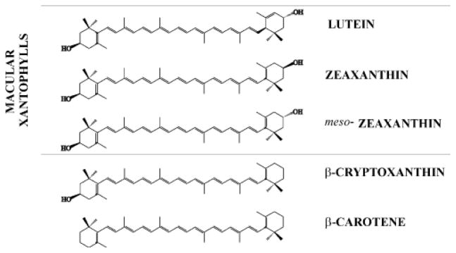
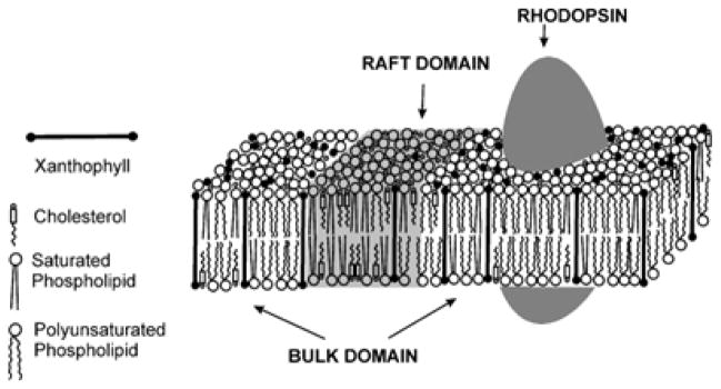
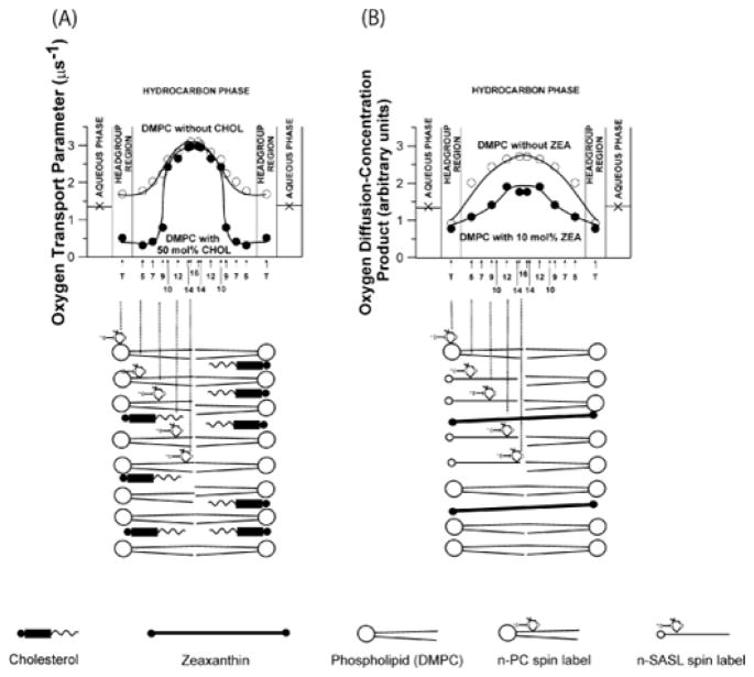
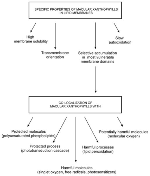
Age-related macular degeneration (AMD) is associated with a low level of macular carotenoids in the eye retina. Only two carotenoids, namely lutein and zeaxanthin are selectively accumulated in the human eye retina from blood plasma where more than twenty other carotenoids are available. The third carotenoid which is found in the human retina, meso-zeaxanthin is formed directly in the retina from lutein. All these carotenoids, named also macular xanthophylls, play key roles in eye health and retinal disease. Macular xanthophylls are thought to combat light-induced damage mediated by reactive oxygen species by absorbing the most damaging incoming wavelength of light prior to the formation of reactive oxygen species (a function expected of carotenoids in nerve fibers) and by chemically and physically quenching reactive oxygen species once they are formed (a function expected of carotenoids in photoreceptor outer segments). There are two major hypotheses about the precise location of macular xanthophylls in the nerve fiber layer of photoreceptor axons and in photoreceptor outer segments. According to the first, macular xanthophylls transversely incorporate in the lipid-bilayer portion of membranes of the human retina. According to the second, macular xanthophylls are protein-bound by membrane-associated, xanthophyll-binding proteins. In this review we indicate specific properties of macular xanthophylls that could help explain their selective accumulation in the primate retina with special attention paid to xanthophyll-membrane interactions.
Keywords: AMD; Carotenoid; Lipid bilayer; Lutein; Macular xanthophylls; Membrane domain; Zeaxanthin.
Figures
References
-
- Britton G, Liaaen-Jensen S, Pfander H. Carotenoids: Handbook. Springer; Basel, Switzerland: 2004.
-
- Khachik F, Beecher GR, Goli MB, Lusby WR. Separation, identification, and quantification of carotenoids in fruits, vegetables and human plasma by high performance liquid chromatography. Pure Appl Chem. 1991;63:71–80.
-
- Khachik F, Spangler CJ, Smith JC, Jr, Canfield LM, Steck A, et al. Identification, quantification, and relative concentrations of carotenoids and their metabolites in human milk and serum. Anal Chem. 1997;69:1873–1881. - PubMed
-
- Bone RA, Landrum JT, Hime GW, Cains A, Zamor J. Stereochemistry of the human macular carotenoids. Invest Ophthalmol Vis Sci. 1993;34:2033–2040. - PubMed
-
- Bone RA, Landrum JT, Friedes LM, Gomez CM, Kilburn MD, et al. Distribution of lutein and zeaxanthin stereoisomers in the human retina. Exp Eye Res. 1997;64:211–218. - PubMed
Grants and funding
LinkOut - more resources
Full Text Sources
Other Literature Sources
Medical