

Immunohistochemical analysis of IL-6, IL-8/CXCR2 axis, Tyr p-STAT-3, and SOCS-3 in lymph nodes from patients with chronic lymphocytic leukemia: correlation between microvascular characteristics and prognostic significance
- PMID: 24883303
- PMCID: PMC4026921
- DOI: 10.1155/2014/251479
Immunohistochemical analysis of IL-6, IL-8/CXCR2 axis, Tyr p-STAT-3, and SOCS-3 in lymph nodes from patients with chronic lymphocytic leukemia: correlation between microvascular characteristics and prognostic significance
Abstract
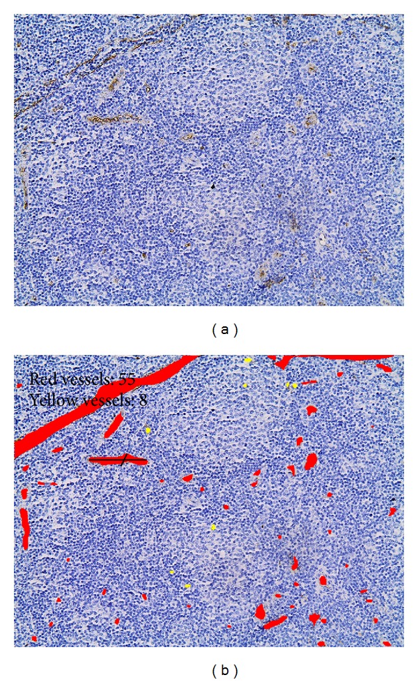
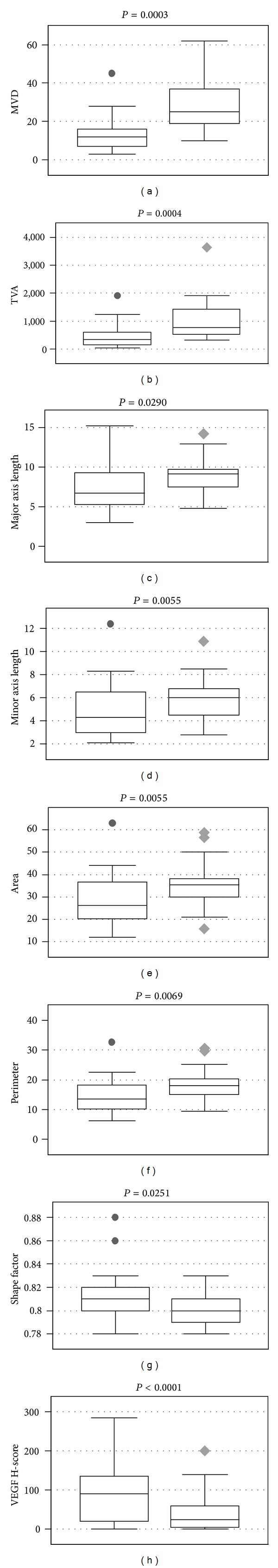
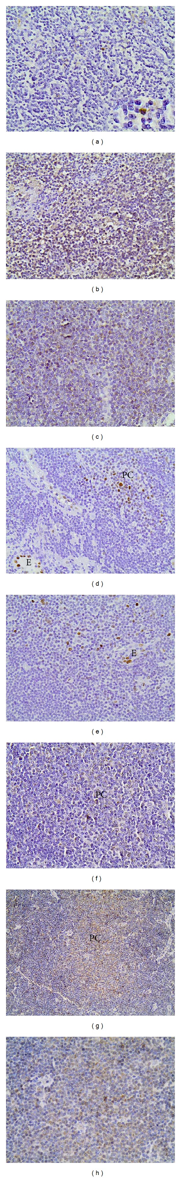
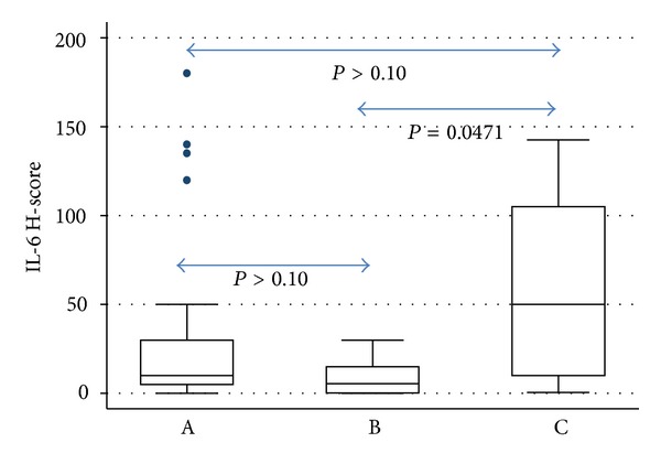
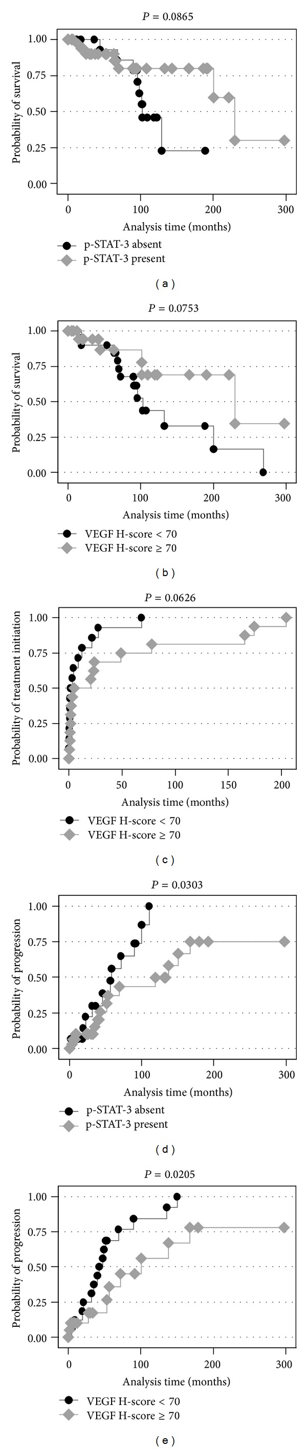
A number of studies have looked into the pathophysiological role of angiogenesis in CLL, but the results have often been inconsistent. We aimed to gain direct insight into the angiogenic process in lymph nodes involved by CLL, focusing on proangiogenic cytokines and microvessel morphometry. The tissue levels of VEGF, Th-2 cytokines IL-6 and IL-8, IL-8 receptor CXCR2, and tyrosine p-STAT-3/SOCS-3 axis modulating cytokine expression were evaluated immunohistochemically in 62 CLL/SLL cases. Microvascular characteristics were evaluated by image analysis. Results were analyzed with regard to clinicopathological characteristics. Proliferation centers (PCs) were less well vascularised compared to non-PC areas. IL-8 and CXCR2 expression was distinctly uncommon as opposed to IL-6, VEGF and SOCS-3, which were detected in the vast majority of cases. The latter two molecule expressions were more pronounced in the PCs in ∼ 40% of the cases. p-STAT-3 immunoreactivity was recorded in 66.67% of the cases with a predilection for PCs. Microvessel morphometry was unrelated to proangiogenic cytokines, p-STAT-3, SOCS-3, or survival. Microvascular caliber and VEGF expression were higher in Binet stage A, whereas IL-6 expression was higher in stage C. VEGF and p-STAT-3 exerted a favorable effect on progression, which remained significant in multivariate analysis, thereby constituting potential outcome predictors in CLL patients.
Figures
Similar articles
-
Expression of interleukin-8 receptor CXCR2 and suppressor of cytokine signaling-3 in astrocytic tumors.Mol Med. 2012 May 9;18(1):379-88. doi: 10.2119/molmed.2011.00449. Mol Med. 2012. PMID: 22231733 Free PMC article.
-
Prognostic significance of IL-8-STAT-3 pathway in astrocytomas: correlation with IL-6, VEGF and microvessel morphometry.Cytokine. 2011 Sep;55(3):387-95. doi: 10.1016/j.cyto.2011.05.012. Cytokine. 2011. PMID: 21684758
-
The role of CXC-chemokine receptor CXCR2 and suppressor of cytokine signaling-3 (SOCS-3) in renal cell carcinoma.BMC Cancer. 2014 Mar 4;14:149. doi: 10.1186/1471-2407-14-149. BMC Cancer. 2014. PMID: 24593195 Free PMC article.
-
The clinical and biologic importance of neovascularization and angiogenic signaling pathways in chronic lymphocytic leukemia.Semin Oncol. 2006 Apr;33(2):174-85. doi: 10.1053/j.seminoncol.2006.01.008. Semin Oncol. 2006. PMID: 16616064 Review.
-
Can the protective actions of JAK-STAT in the heart be exploited therapeutically? Parsing the regulation of interleukin-6-type cytokine signaling.J Cardiovasc Pharmacol. 2007 Aug;50(2):126-41. doi: 10.1097/FJC.0b013e318068dd49. J Cardiovasc Pharmacol. 2007. PMID: 17703129 Review.
Cited by
-
In Chronic Lymphocytic Leukemia the JAK2/STAT3 Pathway Is Constitutively Activated and Its Inhibition Leads to CLL Cell Death Unaffected by the Protective Bone Marrow Microenvironment.Cancers (Basel). 2019 Dec 4;11(12):1939. doi: 10.3390/cancers11121939. Cancers (Basel). 2019. PMID: 31817171 Free PMC article.
-
Hsp90 inhibition increases SOCS3 transcript and regulates migration and cell death in chronic lymphocytic leukemia.Oncotarget. 2016 May 10;7(19):28684-96. doi: 10.18632/oncotarget.8760. Oncotarget. 2016. PMID: 27107422 Free PMC article.
-
CXCR Family and Hematologic Malignancies in the Bone Marrow Microenvironment.Biomolecules. 2025 May 13;15(5):716. doi: 10.3390/biom15050716. Biomolecules. 2025. PMID: 40427609 Free PMC article. Review.
-
Clinico-Biological Implications of Modified Levels of Cytokines in Chronic Lymphocytic Leukemia: A Possible Therapeutic Role.Cancers (Basel). 2020 Feb 24;12(2):524. doi: 10.3390/cancers12020524. Cancers (Basel). 2020. PMID: 32102441 Free PMC article. Review.
-
STAT3 genetic variant, alone and in combination with STAT5b polymorphism, contributes to breast cancer risk and clinical outcomes.Med Oncol. 2015 Jan;32(1):375. doi: 10.1007/s12032-014-0375-z. Epub 2014 Dec 7. Med Oncol. 2015. PMID: 25487443
References
-
- Bergers G, Benjamin LE. Tumorigenesis and the angiogenic switch. Nature Reviews Cancer. 2003;3(6):401–410. - PubMed
-
- Sachanas S, Angelopoulou M, Korkolopoulou Kalpadakis P, et al. Angiogenesis in chronic lymphocytic leukemia. Current Angiogenesis. 2013;2(2):96–103.
-
- Morais Borges N, Colleoni GWB. Angiogenesis and AngiomiRs in Non-Hodgkin’s lymphomas. Current Angiogenesis. 2012;1(1):20–25.
-
- Molica S. Angiogenesis in B-cell chronic lymphocytic leukemia: methods of study, clinical significance and prognostic implications. Leukemia & Lymphoma. 2001;42(4):603–607. - PubMed
-
- Kay NE, Bone ND, Tschumper RC, et al. B-CLL cells are capable of synthesis and secretion of both pro- and anti-angiogenic molecules. Leukemia. 2002;16(5):911–919. - PubMed
MeSH terms
Substances
LinkOut - more resources
Full Text Sources
Other Literature Sources
Miscellaneous
