. 2014 May 17:13:110.
doi: 10.1186/1476-4598-13-110.

Fbxw7 is an independent prognostic marker and induces apoptosis and growth arrest by regulating YAP abundance in hepatocellular carcinoma

Affiliations

Fbxw7 is an independent prognostic marker and induces apoptosis and growth arrest by regulating YAP abundance in hepatocellular carcinoma

Kangsheng Tu et al. Mol Cancer. .

Abstract

Background: The E3 ubiquitin ligase Fbxw7 functions as a general tumor suppressor by targeting several well-known oncoproteins for ubiquitination and proteasomal degradation. However, the clinical significance of Fbxw7 and the mechanisms involved in the anti-cancer effect of Fbxw7 in HCC are not clear.

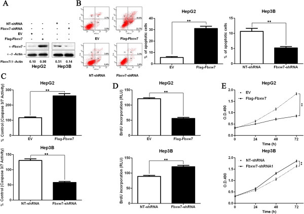
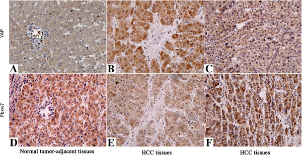
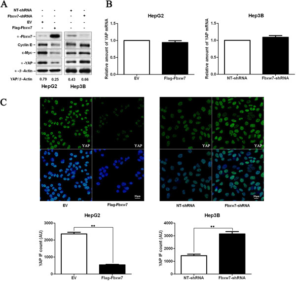
Method: The Fbxw7 and YAP expression in 60 samples of surgical resected HCC and matched normal tumor-adjacent tissues were assessed using IHC or immunoblotting. Flow cytometry, caspase 3/7 activity assay, BrdU cell proliferation assay and MTT assay were used to detect proliferation and apoptosis of HCC cells. The regulatory effect of Fbxw7 on YAP in HCC cells was confirmed by qRT-PCR, immunoblotting and immunofluorescence. Co-immunoprecipitation was used to analyze interaction between YAP and Fbxw7. Nude mice subcutaneous injection, Ki-67 staining and TUNEL assay were used to evaluate tumor growth and apoptosis in vivo.

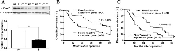
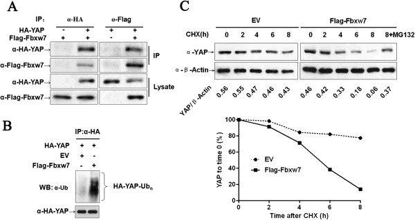
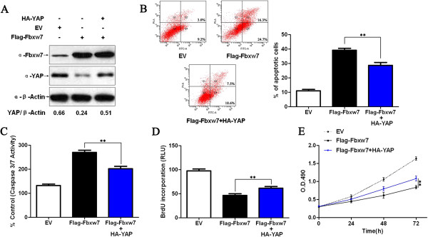
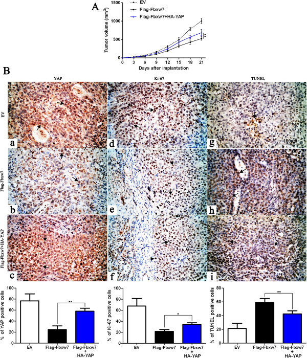
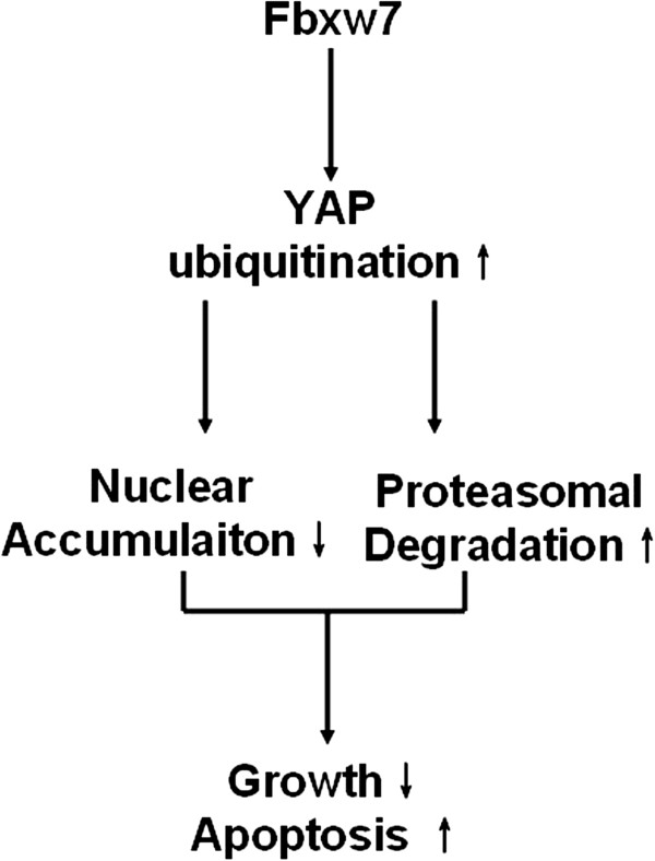
Results: In this study, we found that Fbxw7 expression was impaired in HCC tissues and loss of Fbxw7 expression was correlated with poor clinicopathological features including large tumor size, venous infiltration, high pathological grading and advanced TNM stage. Additionally, we demonstrated that patients with positive Fbxw7 expression had a better 5-year survival and Fbxw7 was an independent factor for predicting the prognosis of HCC patients. We confirmed that Fbxw7 inhibited HCC by inducing both apoptosis and growth arrest. Elevated YAP expression was observed in the same cohort of HCC tissues. Pearson's correlation coefficient analysis indicated that Fbxw7 was inversely associated with YAP protein expression in HCC tissues. We also found that Fbxw7 regulated YAP protein abundance by targeting YAP for ubiquitination and proteasomal degradation in HCC. Furthermore, restoring YAP expression partially abrogated Fbxw7 induced HCC cell apoptosis and growth arrest in vitro and in vivo.

Conclusion: These results indicate that Fbxw7 may serve as a prognostic marker and that YAP may be a potential target of Fbxw7 in HCC.


Figures

Figure 1
Figure 1
Expression of Fbxw7 and its clinical significance in HCC cases. A) Representative western blot analysis of Fbxw7 expression in cancer (T) and matched noncancerous tissues (NT) was shown. Quantification of the data revealed that Fbxw7 protein expression in HCC tissues was significantly lower than that in the normal tumor-adjacent tissues. n = 20; Values are depicted as Mean ± SEM; **P < 0.01 by t test. B) Kaplan–Meier overall 5-year and C) disease-free survival curves for HCC patients according to their Fbxw7 protein expression status. The Fbxw7 positive expression group (n = 24), IHC score of Fbxw7 = 1–3; Fbxw7 negative expression group (n = 36), IHC score of Fbxw7 = 0; *P < 0.05 by log-rank test.
Figure 2
Figure 2
Fbxw7 regulates apoptosis and proliferation in HCC cells. A) HepG2 and Hep3B cells that had been transfected with Flag-Fbxw7 and Fbxw7-shRNA, respectively, were subjected to western blotting for Fbxw7. The data are representative of multiple repeats with similar results. B) Quantification of the apoptotic cell population by flow cytometry. Fbxw7 overexpressing HepG2 cells were composed of a larger subset of apoptotic cells compared with the control cells and Fbxw7 knockdown decreased the percentage of apoptotic Hep3B cells. **P < 0.01 by t test; n = 3 repeats with similar results. C) The activity of the pro-apoptotic caspases 3 and 7 was up-regulated after Fbxw7 overexpression in HepG2 cells and down-regulated after Fbxw7 knockdown in Hep3B cells. **P < 0.01 by t test; n = 3 repeats with similar results. D) Cell proliferation as measured by BrdU incorporation was inhibited by Fbxw7 overexpression in HepG2 cells and promoted by Fbxw7 knockdown in Hep3B cells. **P < 0.01 by t test; n = 3 repeats with similar results. E) As assessed by MTT assays, Fbxw7 overexpression was found to reduce the viability of HepG2 cells and Fbxw7 knockdown was found to enhance the viability of Hep3B cells. **P < 0.01 by two-way ANOVA; n = 3 repeats with similar results. Values are depicted as Mean ± SEM.
Figure 3
Figure 3
Immunohistochemical analyses of YAP and its correlation with Fbxw7 protein in HCC. In cases of high Fbxw7 protein expression (D, F), there was no detectable YAP protein expression (A, C) in the same tissue section. In contrast, in the case of low Fbxw7 protein expression (E), there was strong YAP protein expression (B). The fibrotic septa (B, E) simply represent fibrotic tumor stroma with normal background liver. Scale bar: 100 μm.
Figure 4
Figure 4
Fbxw7 regulates the stability of the YAP protein in HCC cells. A) HepG2 and Hep3B cells that were transfected with Flag-Fbxw7 and Fbxw7-shRNA, respectively, and subjected to Western blotting for Fbxw7, c-Myc, Cyclin E and YAP. Fbxw7 overexpression decreased c-Myc, Cyclin E and YAP protein levels in HepG2 cells, whereas Fbxw7 knockdown led to c-Myc, Cyclin E and YAP accumulation in Hep3B cells. Data are representative of multiple repeats with similar results. B) HepG2 cells transfected with EV or Flag-Fbxw7 and Hep3B cells transfected with non-targeting (NT)-shRNA or Fbxw7-shRNA were harvested for RNA extraction and real time RT-PCR. Fbxw7 overexpression or knockdown did not change YAP mRNA levels. n = 3 independent experiment; Values are depicted as the Mean ± SEM. C) HCC cells that were treated as above were subjected to IF for YAP. Quantification of YAP immunofluorescence revealed that the average level of YAP in the control cells was significantly higher than that in the Fbxw7 overexpressing HepG2 cells and lower than that in the Fbxw7 shRNA transfected Hep3B cells. Scale bar: 20 μm, n = 6; Values are depicted as the Mean ± SEM; **P < 0.01 by t test.
Figure 5
Figure 5
Fbxw7 binds to YAP and promotes the ubiquitin-mediated proteolysis of YAP. A) YAP and Fbxw7 coimmunoprecipitate with each other. Flag-Fbxw7 and HA-YAP plasmids were transfected into HEK293 cells. YAP or Fbxw7 was immunoprecipitated with anti-HA or anti-Flag antibody. Western blotting was performed to detect the specific proteins indicated on the left side of each panel. B) HA-YAP was precipitated from HA-YAP overexpressing HepG2 cells by immunoprecipitation using an anti-HA antibody; YAP ubiquitination was detected by western blotting. Fbxw7 overexpression markedly promoted YAP ubiquitination. C) The Fbxw7 turnover rate was shorter in Fbxw7 overexpressing HepG2 cells. The protein half-life of YAP was analyzed following treatment with cycloheximide. The YAP band intensity was normalized to GAPDH and then normalized to t = 100 controls. The half-life of YAP in Flag-Fbxw7 = 5.2 h (R2 = 0.96) and in EV = 16.5 h (R2 = 0.92). Additionally, treatment with MG132 (a proteasome inhibitor) inhibited Fbxw7 induced YAP degradation in HepG2 cells. The data are representative of multiple independent experiments.
Figure 6
Figure 6
Fbxw7’s suppression of Hep3B cell growth was partially reverted by YAP. A) Flag-Fbxw7 transfected Hep3B cells successfully up-regulated Fbxw7 protein expression as shown by western blot. Fbxw7 overexpression in the same cell line could reduce the levels of YAP. Fbxw7 over-expressing cells that were transfected with HA-YAP partially rescued the phenotype, showing higher YAP levels. The data are representative of multiple repeats with similar results. B) Apoptotic cells were measured by flow cytometry. Restoring YAP expression decreased the percentage of apoptotic cells in Flag-Fbxw7 transfected Hep3B cells. **P < 0.01 by one-way ANOVA; n = 3 repeats with similar results. C) The activity of the pro-apoptotic caspases 3 and 7 in Fbxw7 overexpressing Hep3B cells was decreased by HA-YAP transfection. **P < 0.01 by one-way ANOVA; n = 3 repeats with similar results. D) A BrdU assay showed that YAP promotes proliferation in Fbxw7 overexpressing Hep3B cells. **P < 0.01 by one-way ANOVA; n = 3 repeats with similar results. E) YAP was found to enhance the viability of Fbxw7 over-expressing Hep3B cells (MTT assay). **P < 0.01 by two-way ANOVA; n = 3 repeats with similar results. Values are depicted as the Mean ± SEM.
Figure 7
Figure 7
YAP partially abolishes Fbxw7’s suppression of tumor growth. A) Control Hep3B cells (EV, n = 6), Fbxw7 overexpressing Hep3B cells (Flag-Fbxw7, n = 6) and co-expressing Hep3B cells (Flag-Fbxw7 + HA-YAP, n = 6), respectively, were implanted into nude mice via subcutaneous injection. Tumor nodules were measured using a caliper at different times after implantation. Fbxw7 overexpressing Hep3B cells exhibited a greater tumor-inhibiting effect compared with control cells; however, restoring YAP expression accelerated tumor growth, compared with the Flag-Fbxw7 group. **P < 0.01 by two-way ANOVA. B) Tumor nodules were subjected to immunohistochemical staining for YAP and Ki-67, TUNEL assays and quantitative analysis. Representative immunostaining and TUNEL assays revealed that Fbxw7 overexpression significantly reduced the number of YAP and Ki-67 positive cells and increased the number of apoptotic cells. However, the percentage of YAP and Ki-67 positive cells in tumors arising from the Flag-Fbxw7 + HA-YAP group was significantly higher than that in the tumors from the Flag-Fbxw7 group and the percentage of apoptotic cells in the Flag-Fbxw7 + HA-YAP group was significantly lower than that in the Flag-Fbxw7 group. Black arrows indicate positive cells in each photomicrograph. Scale bar: 100 μm; n = 6; Values are depicted as the Mean ± SEM; *P < 0.05 and **P < 0.01 by one-way ANOVA.
Figure 8
Figure 8
Working model for the tumor suppressive function of Fbxw7 and its downstream pathway. Fbxw7 promotes both apoptosis and the growth arrest of HCC through promoting YAP ubiquitination and proteasomal degradation.

Similar articles

Cited by

References

    1. Wang Z, Inuzuka H, Zhong J, Wan L, Fukushima H, Sarkar FH, Wei W. Tumor suppressor functions of FBW7 in cancer development and progression. FEBS Lett. 2012;586:1409–1418. doi: 10.1016/j.febslet.2012.03.017. - DOI - PMC - PubMed
    1. Welcker M, Orian A, Jin J, Grim JE, Harper JW, Eisenman RN, Clurman BE. The Fbw7 tumor suppressor regulates glycogen synthase kinase 3 phosphorylation-dependent c-Myc protein degradation. Proc Natl Acad Sci U S A. 2004;101:9085–9090. doi: 10.1073/pnas.0402770101. - DOI - PMC - PubMed
    1. Koepp DM, Schaefer LK, Ye X, Keyomarsi K, Chu C, Harper JW, Elledge SJ. Phosphorylation-dependent ubiquitination of cyclin E by the SCFFbw7 ubiquitin ligase. Science. 2001;294:173–177. doi: 10.1126/science.1065203. - DOI - PubMed
    1. Nateri AS, Riera-Sans L, Da Costa C, Behrens A. The ubiquitin ligase SCFFbw7 antagonizes apoptotic JNK signaling. Science. 2004;303:1374–1378. doi: 10.1126/science.1092880. - DOI - PubMed
    1. Tu K, Zheng X, Yin G, Zan X, Yao Y, Liu Q. Evaluation of Fbxw7 expression and its correlation with expression of SREBP-1 in a mouse model of NAFLD. Mol Med Rep. 2012;6:525–530. - PubMed

Publication types

MeSH terms