

Whole body magnetic resonance angiography and computed tomography angiography in the vascular mapping of head and neck: an intraindividual comparison
- PMID: 24884580
- PMCID: PMC4028100
- DOI: 10.1186/1746-160X-10-16
Whole body magnetic resonance angiography and computed tomography angiography in the vascular mapping of head and neck: an intraindividual comparison
Abstract
Introduction: The aim of the study was to compare the detectability of neck vessels with contrast enhanced magnetic resonance angiography (MRA) in the setting of a whole-body MRA and multislice computed tomography angiography (CTA) for preoperative vascular mapping of head and neck.
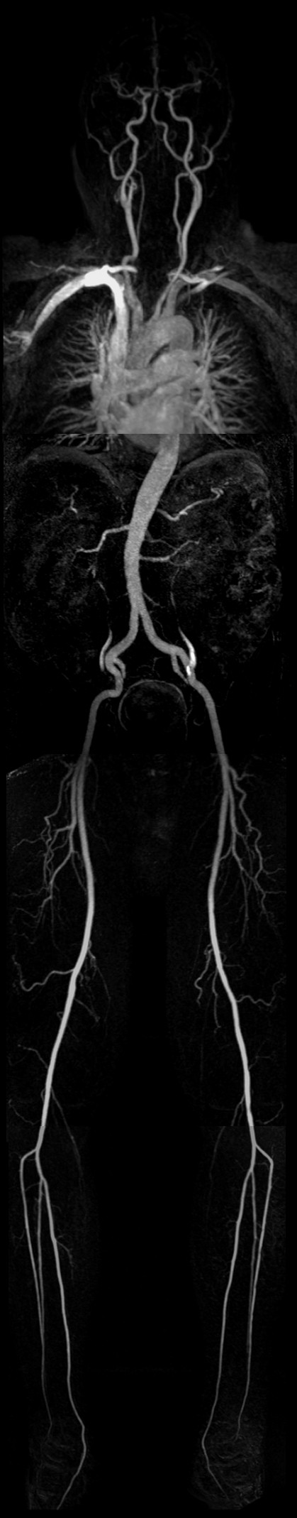
Methods: In 20 patients MRA was performed prior to microvascular reconstruction of the mandible with osteomyocutaneous flaps. CTA of the neck served as the method of reference.1.5 T contrast enhanced magnetic resonance angiograms were acquired to visualize the vascular structures of the neck in the setting of a whole-body MRA examination. 64-slice spiral computed tomography was performed with a dual-phase protocol, using the arterial phase images for 3D CTA reconstruction. Maximum intensity projection was employed to visualize MRA and CTA data. To retrieve differences in the detectability of vessel branches between MRA and CTA, a McNemar test was performed.
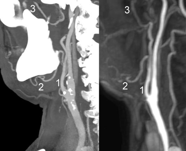
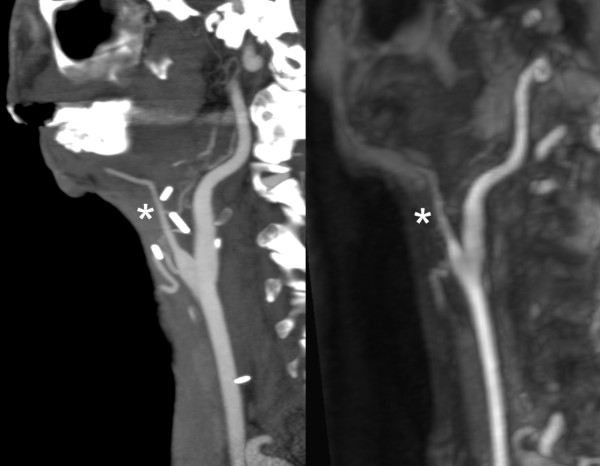
Results: All angiograms were of diagnostic quality. There were no statistically significant differences between MRA and CTA for the detection of branches of the external carotid artery that are relevant host vessels for microsurgery (p = 0.118). CTA was superior to MRA if all the external carotid artery branches were included (p < 0.001).
Conclusions: MRA is a reliable alternative to CTA in vascular mapping of the cervical vasculature for planning of microvascular reconstruction of the mandible. In the setting of whole-body MRA it could serve as a radiation free one-stop-shop tool for preoperative assessment of the arterial system, potentially covering both, the donor and host site in one single examination.
Figures
Similar articles
-
Vascular mapping of head and neck: computed tomography angiography versus digital subtraction angiography.J Oral Maxillofac Surg. 2008 Feb;66(2):302-7. doi: 10.1016/j.joms.2007.05.031. J Oral Maxillofac Surg. 2008. PMID: 18201613
-
Computed tomography angiography versus digital subtraction angiography in vascular mapping for planning of microsurgical reconstruction of the mandible.Eur Radiol. 2005 Aug;15(8):1514-20. doi: 10.1007/s00330-005-2770-5. Epub 2005 Apr 27. Eur Radiol. 2005. PMID: 15856243
-
3 T contrast-enhanced magnetic resonance angiography for evaluation of the intracranial arteries: comparison with time-of-flight magnetic resonance angiography and multislice computed tomography angiography.Invest Radiol. 2006 Nov;41(11):799-805. doi: 10.1097/01.rli.0000242835.00032.f5. Invest Radiol. 2006. PMID: 17035870 Clinical Trial.
-
Vascular imaging of the head and neck.Semin Neurol. 2012 Sep;32(4):401-10. doi: 10.1055/s-0032-1331811. Epub 2013 Jan 29. Semin Neurol. 2012. PMID: 23361484 Review.
-
Value of CTA/MRA in the setting of intraparenchymal hemorrhage in the emergency department.Neuroradiology. 2023 Jan;65(1):97-103. doi: 10.1007/s00234-022-03080-y. Epub 2022 Nov 17. Neuroradiology. 2023. PMID: 36385589 Review.
Cited by
-
Dose reduction potential of iterative reconstruction algorithms in neck CTA-a simulation study.Dentomaxillofac Radiol. 2016 Oct;45(8):20160228. doi: 10.1259/dmfr.20160228. Epub 2016 Aug 19. Dentomaxillofac Radiol. 2016. PMID: 27461784 Free PMC article.
-
The evolving role of MRI in dentomaxillofacial diagnostics: a comprehensive review.Eur Oral Res. 2025 Jan 5;59(1):58-67. doi: 10.26650/eor.2024145664. Eur Oral Res. 2025. PMID: 40453404 Free PMC article. Review.
-
Using 3D printed models for planning and guidance during endovascular intervention: a technical advance.Diagn Interv Radiol. 2015 Jul-Aug;21(4):338-41. doi: 10.5152/dir.2015.14469. Diagn Interv Radiol. 2015. PMID: 26027767 Free PMC article.
-
Salvage para-aortic lymphadenectomy in recurrent cervical cancer after visualization with 3-dimensional computed tomography angiography.Obstet Gynecol Sci. 2018 Sep;61(5):626-630. doi: 10.5468/ogs.2018.61.5.626. Epub 2018 Aug 6. Obstet Gynecol Sci. 2018. PMID: 30255000 Free PMC article.
-
Non-contrast-enhanced magnetic resonance angiography of facial arteries for pre-operative evaluation of vascularized submental lymph node flaps.BMC Med Imaging. 2019 Aug 16;19(1):68. doi: 10.1186/s12880-019-0368-7. BMC Med Imaging. 2019. PMID: 31420022 Free PMC article.
References
-
- Ehrenfeld M, Riediger D, Wolburg H, Thron A. Angiographic visualization and morphology of anastomosed vessels in microsurgical tissue transplantation. Fortschr Kiefer Gesichtschir. 1987;32:71–74. - PubMed
-
- Thurmuller P, Kesting MR, Holzle F, Retzgen H, Wolff KD. Volume-rendered three-dimensional spiral computed tomographic angiography as a planning tool for microsurgical reconstruction in patients who have had operations or radiotherapy for oropharyngeal cancer. Br J Oral Maxillofac Surg. 2007;45:543–547. doi: 10.1016/j.bjoms.2007.03.004. - DOI - PubMed
MeSH terms
LinkOut - more resources
Full Text Sources
Other Literature Sources
Medical
