

Review
doi: 10.1186/1741-7007-12-35.
Shaping the dynamic mitochondrial network
Affiliations
- PMID: 24884775
- PMCID: PMC4035697
- DOI: 10.1186/1741-7007-12-35
Item in Clipboard
Review
Shaping the dynamic mitochondrial network
BMC Biol.
.
Abstract
In a majority of cell types, mitochondria form highly dynamic, tubular networks. Maintaining the shape of this complex network is critical for both mitochondrial and cellular function and involves the activities of mitochondrial division, fusion, motility, and tethering. Recent studies have advanced our understanding of the molecular mechanisms underlying these conserved activities and their integration with cellular needs.
Figures
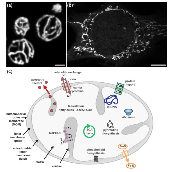
The form and functions of mitochondria. (a) In yeast, mitochondria form a connected, tubular network that is evenly distributed at the cell cortex. (b) Mitochondria also form well-distributed tubular networks in a majority of mammalian cell types. The mitochondrial network of a mouse embryonic fibroblast is shown. Scale bar, 2 μ for (a,b). (c) Like their bacterial ancestors, mitochondria possess two structurally and functionally distinct membranes, the mitochondrial outer and inner membranes (MOM and MIM, respectively). The MOM and MIM surround two compartments, the inner membrane space and matrix, respectively. The matrix houses the circular mitochondrial genome (mtDNA), which encodes protein components of the respiratory complexes I to IV. The MIM, the most protein dense membrane in the cell, adopts elaborate folds called cristae in which assembled respiratory complexes are housed. In addition to ATP production via oxidative phosphorylation, mitochondria play critical roles in phospholipid biosynthesis, metabolite exchange/buffering, β-oxidation of fatty acids, iron-sulfur cluster biogenesis, pyrimidine biosynthesis and the storage and release of apoptotic factors (reviewed in [1]). TCA, tricarboxylic acid.
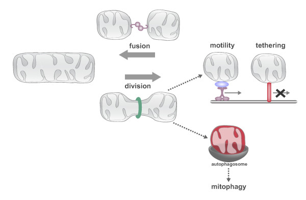
The conserved activities of mitochondrial division, fusion, motility and tethering shape and position the dynamic mitochondrial network. The connectivity of the mitochondrial network is controlled by the antagonistic activities of mitochondrial division and fusion. Mitochondrial division and fusion serve to create a compartment that is a connected conductor, able to mix its contents and have access to mtDNA and its products, but able to be distributed to distant cellular destinations via motor-dependent transport on actin or microtubule networks. Once transported to areas of demand, tethers ensure mitochondria are retained at these cellular locations. In addition to creating transportable mitochondrial compartments, mitochondrial division can produce functionally asymmetric daughter mitochondria. Dysfunctional daughters (depicted in red) cannot re-fuse with the network and are flagged for autophagic degradation.
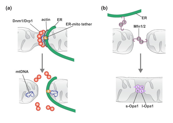
Molecular models of mitochondrial division and fusion. (a) Mitochondrial division involves communication between extra-mitochondrial division factors and internal mitochondrial structures. See text for details. (b) Mitochondrial fusion requires the sequential interaction of the MOM and MIM. MOM fusion is mediated by Mfn1/2, and MIM fusion is mediated by Opa1. Mfn2 is also localized to the ER and functions to tether the ER and mitochondria.
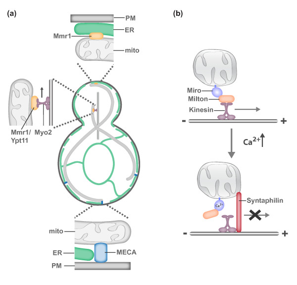
Molecular models of mitochondrial motility and tethering. (a) In yeast, mitochondria are actively transported to the growing bud via Myo2-driven transport along the actin cytoskeleton. Myo2-driven transport requires either Mmr1 or Ypt11 [76-82]. Mother- and bud-specific mitochondrial tethers ensure that both cells retain part of the essential mitochondrial compartment. The mother specific tether MECA (mitochondria-ER-cortex anchor) is composed of three membranes, the plasma membrane (PM), ER and mitochondria, and at least two proteins, Num1 and Mdm36 [84]. In addition to serving as a mitochondrial adaptor for Myo2, Mmr1 functions to tether mitochondria to ER sheets at the bud tip [83]. (b) A model for activity-dependent transport and tethering of mitochondria in axons. The conserved MOM Rho-like GTPase Miro and its binding partner Milton function as a mitochondrial receptor for kinesin. In active synaptic regions, Ca2+-binding by Miro triggers a confirmation change that disrupts kinesin-driven mitochondrial transport [98,99]. Ca2+-mediated confirmation changes have been proposed to disrupt the interaction between Miro/Milton with kinesin (shown here) or kinesin with microtubules. In response to neuronal activity (elevated Ca2+), syntaphilin is also recruited to mitochondria and functions as a static mitochondria-microtubule tether [100].
References
Publication types
MeSH terms
LinkOut - more resources
Full Text Sources
Other Literature Sources
Molecular Biology Databases
