

Sequence artefacts in a prospective series of formalin-fixed tumours tested for mutations in hotspot regions by massively parallel sequencing
- PMID: 24885028
- PMCID: PMC4032349
- DOI: 10.1186/1755-8794-7-23
Sequence artefacts in a prospective series of formalin-fixed tumours tested for mutations in hotspot regions by massively parallel sequencing
Abstract
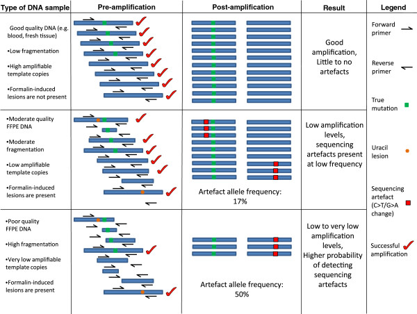
Background: Clinical specimens undergoing diagnostic molecular pathology testing are fixed in formalin due to the necessity for detailed morphological assessment. However, formalin fixation can cause major issues with molecular testing, as it causes DNA damage such as fragmentation and non-reproducible sequencing artefacts after PCR amplification. In the context of massively parallel sequencing (MPS), distinguishing true low frequency variants from sequencing artefacts remains challenging. The prevalence of formalin-induced DNA damage and its impact on molecular testing and clinical genomics remains poorly understood.
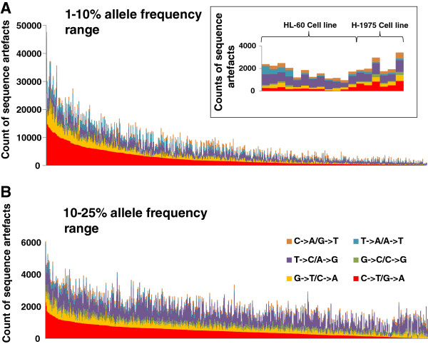
Methods: The Cancer 2015 study is a population-based cancer cohort used to assess the feasibility of mutational screening using MPS in cancer patients from Victoria, Australia. While blocks were formalin-fixed and paraffin-embedded in different anatomical pathology laboratories, they were centrally extracted for DNA utilising the same protocol, and run through the same MPS platform (Illumina TruSeq Amplicon Cancer Panel). The sequencing artefacts in the 1-10% and the 10-25% allele frequency ranges were assessed in 488 formalin-fixed tumours from the pilot phase of the Cancer 2015 cohort. All blocks were less than 2.5 years of age (mean 93 days).
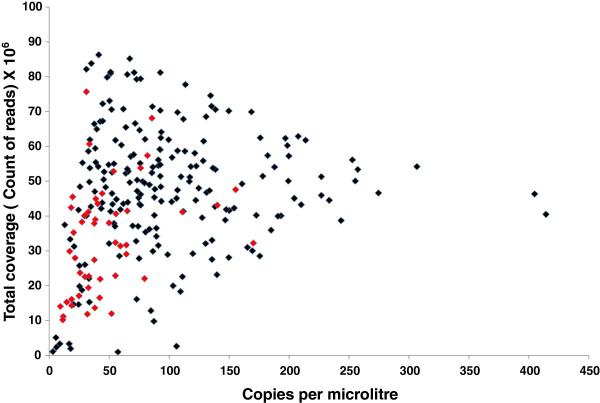
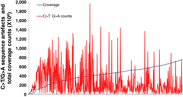
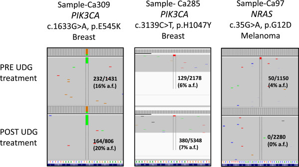
Results: Consistent with the signature of DNA damage due to formalin fixation, many formalin-fixed samples displayed disproportionate levels of C>T/G>A changes in the 1-10% allele frequency range. Artefacts were less apparent in the 10-25% allele frequency range. Significantly, changes were inversely correlated with coverage indicating high levels of sequencing artefacts were associated with samples with low amounts of available amplifiable template due to fragmentation. The degree of fragmentation and sequencing artefacts differed between blocks sourced from different anatomical pathology laboratories. In a limited validation of potentially actionable low frequency mutations, a NRAS G12D mutation in a melanoma was shown to be a false positive.
Conclusions: These findings indicate that DNA damage following formalin fixation remains a major challenge in laboratories working with MPS. Methodologies that assess, minimise or remove formalin-induced DNA damaged templates as part of MPS protocols will aid in the interpretation of genomic results leading to better patient outcomes.
Figures
References
-
- MacConaill LE, Campbell CD, Kehoe SM, Bass AJ, Hatton C, Niu L, Davis M, Yao K, Hanna M, Mondal C, Luongo L, Emery CM, Baker AC, Philips J, Goff DJ, Fiorentino M, Rubin MA, Polyak K, Chan J, Wang Y, Fletcher JA, Santagata S, Corso G, Roviello F, Shivdasani R, Kieran MW, Ligon KL, Stiles CD, Hahn WC, Meyerson ML. et al. Profiling critical cancer gene mutations in clinical tumor samples. PloS one. 2009;7:e7887. doi: 10.1371/journal.pone.0007887. - DOI - PMC - PubMed
-
- Mar VJ, Wong SQ, Li J, Scolyer RA, McLean C, Papenfuss AT, Tothill RW, Kakavand H, Mann GJ, Thompson JF, Behren A, Cebon JS, Wolfe R, Kelly JW, Dobrovic A, McArthur GA. BRAF/NRAS wild-type melanomas have a high mutation load correlating with histologic and molecular signatures of UV damage. Clin Cancer Res. 2013;7:4589–4598. doi: 10.1158/1078-0432.CCR-13-0398. - DOI - PubMed
-
- Wagle N, Berger MF, Davis MJ, Blumenstiel B, Defelice M, Pochanard P, Ducar M, Van Hummelen P, Macconaill LE, Hahn WC, Meyerson M, Gabriel SB, Garraway LA. High-throughput detection of actionable genomic alterations in clinical tumor samples by targeted, massively parallel sequencing. Cancer discovery. 2012;7(1):82–93. doi: 10.1158/2159-8290.CD-11-0184. - DOI - PMC - PubMed
Publication types
MeSH terms
Substances
LinkOut - more resources
Full Text Sources
Other Literature Sources
Miscellaneous
