

Anterior segment dysgenesis associated with Williams-Beuren syndrome: a case report and review of the literature
- PMID: 24885071
- PMCID: PMC4031488
- DOI: 10.1186/1471-2415-14-70
Anterior segment dysgenesis associated with Williams-Beuren syndrome: a case report and review of the literature
Abstract
Background: Williams-Beuren syndrome is characterized by mild mental retardation, specific neurocognitive profile, hypercalcemia during infancy, distinctive facial features and cardiovascular diseases. We report on complete ophthalmologic, sonographic and genetic evaluation of a girl with a clinical phenotype of Williams-Beuren syndrome, associated with unilateral anterior segment dysgenesis and bilateral cleft of the soft and hard palate. These phenotypic features have not been linked to the haploinsufficiency of genes involved in the microdeletion.
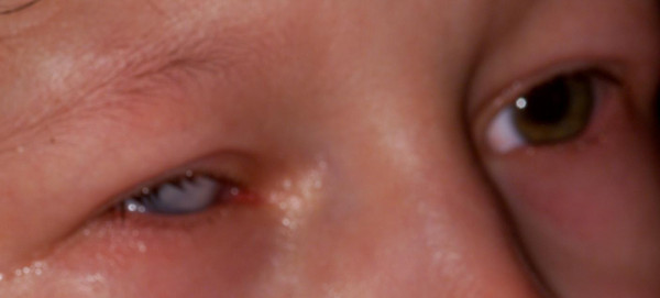
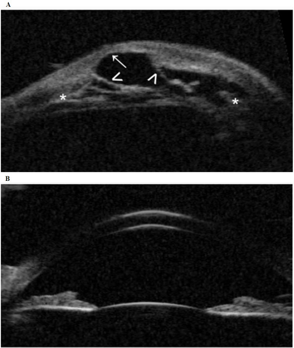
Case presentation: A term born girl presented at the initial examination with clouding of the right cornea. On ultrasound biomicroscopy the anterior chamber structures were difficult to differentiate, showing severe adhesions from the opacified cornea to the iris with a kerato-irido-lenticular contact to the remnant lens, a finding consistent with Peters' anomaly. Genetic analyses including FISH confirmed a loss of the critical region 7q11.23, usually associated with the typical Williams-Beuren syndrome. Microsatellite analysis showed a loss of about 2.36 Mb.
Conclusions: A diagnosis of Williams-Beuren syndrome was made based on the microdeletion of 7q11.23. The unique features, including unilateral microphthalmia and anterior segment dysgenesis, were unlikely to be caused by the microdeletion. Arguments in favor of the latter are unilateral manifestation, as well as the fact that numerous patients with deletions of comparable or microscopically visible size have not shown similar manifestations.
Figures
Similar articles
-
8q21.11 microdeletion in two patients with syndromic peters anomaly.Am J Med Genet A. 2016 Sep;170(9):2471-5. doi: 10.1002/ajmg.a.37840. Epub 2016 Jul 5. Am J Med Genet A. 2016. PMID: 27378168 Free PMC article. Review.
-
Ocular Manifestations of Peters Plus-Like Syndrome in 8q21.11 Microdeletion Syndrome.Cornea. 2023 Jul 1;42(7):908-911. doi: 10.1097/ICO.0000000000003281. Epub 2023 Apr 6. Cornea. 2023. PMID: 37039706
-
Genetics of Congenital Corneal Opacification--Impact on Diagnosis and Treatment.Cornea. 2015 Oct;34 Suppl 10:S24-34. doi: 10.1097/ICO.0000000000000552. Cornea. 2015. PMID: 26352876 Review.
-
Peters Anomaly: Novel Non-Invasive Alternatives to Penetrating Keratoplasty.Semin Ophthalmol. 2023 Apr;38(3):275-282. doi: 10.1080/08820538.2023.2176238. Epub 2023 Feb 14. Semin Ophthalmol. 2023. PMID: 36788651 Review.
-
[Clinical aspects and genetics of Williams-Beuren syndrome. Clinical and molecular genetic study of 44 patients with suspected Williams-Beuren syndrome].Klin Padiatr. 2000 Nov-Dec;212(6):299-307. doi: 10.1055/s-2000-9605. Klin Padiatr. 2000. PMID: 11190824 German.
Cited by
-
Assessment of the anterior segment of patients with primary congenital glaucoma using handheld optical coherence tomography.Eye (Lond). 2019 Aug;33(8):1232-1239. doi: 10.1038/s41433-019-0369-3. Epub 2019 Mar 18. Eye (Lond). 2019. PMID: 30886322 Free PMC article.
-
Anterior pituitary, sex hormones, and keratoconus: Beyond traditional targets.Prog Retin Eye Res. 2022 May;88:101016. doi: 10.1016/j.preteyeres.2021.101016. Epub 2021 Nov 2. Prog Retin Eye Res. 2022. PMID: 34740824 Free PMC article.
References
-
- Beuren AJ. A new syndrome: supravalvular aortic stenosis, multiple peripheral pulmonary stenosis, mental retardation, similar facial features and identical dental abnormalities. Monatsschr Kinderheilkd. 1964;112:218–221. - PubMed
-
- Black JA, Cartier R. Association between aortic stenosis and facies of severe infantile hypercalcaemia. Lancet. 1963;2:745–749. - PubMed
-
- Axelsson S. Variability of the cranial and dental phenotype in Williams syndrome. Swed Dent J Suppl. 2005;170:3–67. - PubMed
Publication types
MeSH terms
Supplementary concepts
LinkOut - more resources
Full Text Sources
Other Literature Sources
Miscellaneous
