. 2014 May 29:11:97.
doi: 10.1186/1742-2094-11-97.

MiR-155 induction in microglial cells suppresses Japanese encephalitis virus replication and negatively modulates innate immune responses

Affiliations

MiR-155 induction in microglial cells suppresses Japanese encephalitis virus replication and negatively modulates innate immune responses

Siddhika Pareek et al. J Neuroinflammation. .

Abstract

Background: Microglial cells, which are resident macrophages of the central nervous system, play important roles in immune responses and pathogenesis. Japanese encephalitis virus (JEV) is a neurotropic virus that infects microglial cells in brain. Several microRNAs including miR-155 and miR-146a play an important role in defining the microglia inflammatory profile. In this study, we have investigated the effect of miR-155 and miR-146a modulation on JEV infection as well as innate immune responses in human microglial cells.

Methods: In vitro studies were performed in JEV-infected human microglial CHME3 cells. miR-155 or miR-146a were overexpressed and total RNA and protein were extracted following JEV-infection. Expression of genes involved in innate immune responses was studied by PCR array, quantitative real-time PCR (qPCR), western blot and Fluorescence activated cell sorter (FACS). JEV replication was monitored by studying the viral RNA by qPCR, protein by western blot, and titres by plaque assay.

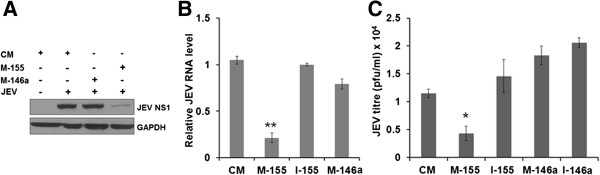
Results: Overexpression of miR-155 in CHME3 cells resulted in significantly reduced JEV replication whereas miR-146a overexpression had an insignificant effect. Additionally, interferon regulatory factor 8 (IRF8) and complement factor H (CFH) were induced during JEV infection; however, this induction was attenuated in miR-155 overexpressing cells following JEV infection. Further, JEV-induced NF-κB regulated downstream gene expression was attenuated. Interestingly, an increased level of CD45, a negative regulator of microglia activation and a reduced phosphorylated-Signal Transducers and Activators of Transcription (p-STAT1) expression was observed in miR-155 overexpressing cells upon JEV infection.

Conclusion: Induction of miR-155 in human microglial cells may negatively modulate JEV-induced innate immune gene expression and may have a beneficial role in limiting JEV replication in human microglial cells.


Figures

Figure 1
Figure 1
Differential expression of NeurimmiRs in human microglial cells after JEV infection. CHME3 cells were mock-infected or infected with JEV. Total RNA was isolated from cells at different time points. (A) Relative abundance of six NeurimmiRs obtained from human miRNA microarray was shown as log2 fold-change. (B) Levels of miR-155 and miR-146a were determined by qPCR and plotted as relative to that seen in the mock-infected cells. Data were normalized against U6 snRNA. h, hours; JEV, Japanese encephalitis virus; pi, post-infection. *, P <0.05; **, P <0.005.
Figure 2
Figure 2
JEV replication in human microglial cells. CHME3 cells were transfected with control mimic (CM), mimic-155 (M-155) and mimic-146a (M-146a) followed by JEV infection. Virus replication was monitored at 24 hours pi. (A) JEV NS1 expression was studied using western blotting. (B) Relative levels of JEV RNA were quantified by qPCR. Data were normalized against GAPDH RNA. (C) JEV titres in culture supernatants were determined by plaque assay. GAPDH, Glyceraldehyde 3-phosphate dehydrogenase; JEV, Japanese encephalitis virus; pi, post-infection. *, P <0.05; **, P <0.005.
Figure 3
Figure 3
JEV-induced IFN-β mRNA expression in human microglial cells. CHME3 cells were transfected with CM, M-155, M-146a, or I-155 and mock-infected or infected with JEV 24 hours later. Total RNA was extracted 24 hours pi. and levels of various transcripts were determined by qPCR normalized against those of GAPDH. (A) Relative IFNβ mRNA expression in JEV-infected cells compared to mock-infected cells. (B) Relative abundance of various ISG transcripts in JEV-infected cells compared to mock-infected cells. (C) Relative expression of IFIT1 in M-155 and I-155 transfected cells compared to CM transfected cells following JEV infection. CM, control mimic; GAPDH, Glyceraldehyde 3-phosphate dehydrogenase; I-155, h, hours; inhibitor 155; IFN, interferon; ISG, interferon-stimulated genes; JEV, Japanese encephalitis virus; M-146a, mimic 146a; M-155, mimic 155; pi, post-infection. **, P <0.005.
Figure 4
Figure 4
JEV-induced IRF8 expression in human microglial cells. (A) CHME3 cells were mock-infected or infected with JEV and total RNA was extracted from cells harvested at different time points. Relative levels of IRF8 and IRF3 transcripts in JEV-infected cells compared to those in mock-infected cells are presented in the left panel. GAPDH expression was used to normalize the data. The right panel shows IRF8 protein levels during JEV infection. (B) CHME3 cells were transfected with CM, M-155 and I-155 followed by JEV infection. The left panel shows the relative abundance of IRF8 measured by qPCR in JEV infected cells in comparison to that in mock-infected cells. GAPDH expression was used to normalize the data. The right panel shows the western blot of IRF8. CM, control mimic; GAPDH, Glyceraldehyde 3-phosphate dehydrogenase; h, hours; I-146a, inhibitor 146a; I-155, inhibitor 155; IRF, interferon regulatory factor; JEV, Japanese encephalitis virus; M-146a, mimic 146a; M-155, mimic 155; pi, post-infection. *, P <0.05.
Figure 5
Figure 5
JEV-induced NF-κB targeted gene expression in human microglial cells. (A) CHME3 cells were treated for 6 hours with NF-κB inhibitor (25 μM) or PI3K inhibitor (10 μM) using DMSO as a vehicle. This was followed by JEV infection for 24 hours. Total RNA was extracted and relative IFN-β mRNA expression was measured using qPCR in inhibitor-treated cells compared to the vehicle-treated cells. GAPDH was used to normalize the data. (B) CHME3 cells were transfected with CM, M-155 or M-146a and infected with JEV 24 hours later. Total RNA was extracted 24 hours pi and levels of various NF-κB targeted genes were studied using PCR array. Hierarchical clustering represents the co-regulated genes across the groups. Relative gene expression levels were depicted according to the colour scale (green - downregulation, red - upregulation). Gene designations are listed to the left. (C) Expression of NF-κB targeted genes expression that were found to be common (n = 19) in PCR array among three different groups (CM + JEV, M-146a + JEV, and M-155 + JEV) in CHME3 cells. An attenuated expression of JEV-induced genes was observed in miR-155 overexpressing cells. (D) Expression of IL-12B, PTGS-2, CCR5 and IL-4 was studied by qPCR. The figure shows a relative abundance of transcripts in JEV-infected, mimic-transfected CHME3 cells when compared with those in cells transfected with the mimics. GAPDH was used to normalize the data. (E) Expression of MyD88 and Ikkϵ in miR-155 or miR-146a overexpressing JEV-infected CHME3 cells as seen by western blotting. (F) Expression of different mRNAs in miR-155 overexpressing CHME3 cells following infection with JEV. CM, control mimic; DMSO, Dimethyl sulfoxide; GAPDH, Glyceraldehyde 3-phosphate dehydrogenase; IFN, interferon; JEV, Japanese encephalitis virus; M-146a, mimic 146a; M-155, mimic 155.
Figure 6
Figure 6
JEV-induced CD45 expressions in human microglial cells. (A) CHME3 cells were transfected with CM, M-155 or I-155 followed by JEV infection for 24 hours. Cells were harvested and examined for CD45 expression by FACS. Data were analyzed by FlowJo software (Tree Star). Y-axis represents the number of events normalized according to FlowJo algorithms. (B)p-STAT1 and STAT1 expression was studied in JEV-infected cells by western blotting. (C) Relative expression of TNF-α, IL-1β, IL-4 and IL10 mRNA in M-155 transfected cells infected with JEV when compared with those transfected with CM. The data were normalized against GAPDH. CM, control mimic; FACS, fluorescence activated cell sorter; GAPDH, Glyceraldehyde 3-phosphate dehydrogenase; I-155, inhibitor 155; JEV, Japanese encephalitis virus; M-146a, mimic 146a; M-155, mimic 155.
Figure 7
Figure 7
JEV-induced CFH expression in human microglial cells. (A) CHME3 cells were mock-infected or infected with JEV. Total RNA was extracted from cells at different time points. The relative mRNA levels of CFH (left panel) and CD59, CD55, and CD46 (right panel) were determined in JEV-infected cells using qPCR in comparison to that in mock-infected cells. GAPDH was used as the normalizing control. (B) CHME3 cells were treated with either DMSO as a vehicle, NF-κB (25 μM) or PI3K inhibitor (10 μM) in DMSO for 6 hours followed by JEV infection for 24 hours. Total RNA was extracted and relative levels of CFH expression was studied by qPCR in different treatments when compared with vehicle-treated cells. (C) CHME3 was transfected with CM, M-155 or M-146a. After 24 hours of transfection cells were either mock-infected or infected with JEV for another 24 hours. Total RNA was extracted and relative levels of CFH mRNA determined in JEV-infected cells in comparison to those seen in mock-infected cells. CFH, complement factor H; CM, control mimic; DMSO, Dimethyl sulfoxide; GAPDH, Glyceraldehyde 3-phosphate dehydrogenase; h, hours; JEV, Japanese encephalitis virus; M-146a, mimic 146a; M-155, mimic 155; pi, post-infection. *, P <0.05; **, P <0.005.

Similar articles

Cited by

References

    1. Thongtan T, Cheepsunthorn P, Chaiworakul V, Rattanarungsan C, Wikan N, Smith DR. Highly permissive infection of microglial cells by Japanese encephalitis virus: a possible role as a viral reservoir. Microbes Infect. 2010;12:37–45. doi: 10.1016/j.micinf.2009.09.013. - DOI - PubMed
    1. Chen CJ, Ou YC, Lin SY, Raung SL, Liao SL, Lai CY, Chen SY, Chen JH. Glial activation involvement in neuronal death by Japanese encephalitis virus infection. J Gen Virol. 2010;91:1028–1037. doi: 10.1099/vir.0.013565-0. - DOI - PubMed
    1. Das S, Dutta K, Kumawat KL, Ghoshal A, Adhya D, Basu A. Abrogated inflammatory response promotes neurogenesis in a murine model of Japanese encephalitis. PLoS One. 2011;6:e17225. doi: 10.1371/journal.pone.0017225. - DOI - PMC - PubMed
    1. Kobayashi K, Imagama S, Ohgomori T, Hirano K, Uchimura K, Sakamoto K, Hirakawa A, Takeuchi H, Suzumura A, Ishiguro N, Kadomatsu K. Minocycline selectively inhibits M1 polarization of microglia. Cell Death Dis. 2013;4:e525. doi: 10.1038/cddis.2013.54. - DOI - PMC - PubMed
    1. Mishra MK, Basu A. Minocycline neuroprotects, reduces microglial activation, inhibits caspase 3 induction, and viral replication following Japanese encephalitis. J Neurochem. 2008;105:1582–1595. doi: 10.1111/j.1471-4159.2008.05238.x. - DOI - PubMed

Publication types

MeSH terms