. 2014 May 2:14:308.
doi: 10.1186/1471-2407-14-308.

Demethylation of the miR-146a promoter by 5-Aza-2'-deoxycytidine correlates with delayed progression of castration-resistant prostate cancer

Affiliations

Demethylation of the miR-146a promoter by 5-Aza-2'-deoxycytidine correlates with delayed progression of castration-resistant prostate cancer

Xiaolu Wang et al. BMC Cancer. .

Abstract

Background: Androgen deprivation therapy is the primary strategy for the treatment of advanced prostate cancer; however, after an initial regression, most patients will inevitably develop a fatal androgen-independent tumor. Therefore, understanding the mechanisms of the transition to androgen independence prostate cancer is critical to identify new ways to treat older patients who are ineligible for conventional chemotherapy.

Methods: The effects of 5-Aza-2'-deoxycytidine (5-Aza-CdR) on the viability and the apoptosis of the androgen-dependent (LNCaP) and androgen-independent (PC3) cell lines were examined by MTS assay and western blot analysis for the activation of caspase-3. The subcutaneous LNCaP xenografts were established in a nude mice model. MiR-146a and DNMTs expressions were analyzed by qRT-PCR and DNA methylation rates of LINE-1 were measured by COBRA-IRS to determine the global DNA methylation levels. The methylation levels of miR-146a promoter region in the different groups were quantified by the bisulfite sequencing PCR (BSP) assay.

Results: We validated that 5-Aza-CdR induced cell death and increased miR-146a expression in both LNCaP and PC3 cells. Notably, the expression of miR-146a in LNCaP cells was much higher than in PC3 cells. MiR-146a inhibitor was shown to suppress apoptosis in 5-Aza-CdR-treated cells. In a castrate mouse LNCaP xenograft model, 5-Aza-CdR significantly suppressed the tumors growth and also inhibited prostate cancer progression. Meanwhile, miR-146a expression was significantly enhanced in the tumor xenografts of 5-Aza-CdR-treated mice and the androgen-dependent but not the androgen-independent stage of castrated mice. In particular, the expression of miR-146a was significantly augmented in both stages of the combined treatment (castration and 5-Aza-CdR). Additionally, the methylation percentage of the two CpG sites (-444 bp and -433 bp), which were around the NF-κB binding site at miR-146a promoter, showed the lowest methylation levels among all CpG sites in the combined treatment tumors of both stages.

Conclusion: Up-regulating miR-146a expression via the hypomethylation of the miR-146a promoter by 5-Aza-CdR was correlated with delayed progression of castration-resistant prostate cancers. Moreover, site-specific DNA methylation may play an important role in miR-146a expression in androgen-dependent prostate cancer progression to androgen-independent prostate cancer and therefore provides a potentially useful biomarker for assessing drug efficacy in prostate cancer.


Figures

Figure 1
Figure 1
The effect of 5-Aza-CdR and miR-146a inhibitors on the viability and apoptosis of LNCaP and PC3 prostate cancer cells. LNCaP and PC3 prostate cancer cells transfected with the miR-146a inhibitor or control inhibitor were treated with increasing doses of 5-aza-CdR, replenished daily for up to 6 days. The relative cell viability was measured by the MTS assay (A-B) and apoptosis was detected by western blot analysis for the activation of caspase-3 (C). MiR-146a expression was measured by qRT-PCR in LNCaP and PC3 cells treated with 5-Aza-CdR at various concentrations for 4 days transfected with control inhibitor or miR-146a inhibitor (D). DNA methylation levels of miR-146a promoter region in 5-Aza-CdR-treated LNCaP cells by BSP assay (E). The data represent the mean ± SD of triplicate wells from at least two experiments. *P < 0.05 and **P < 0.01 compared to the control.
Figure 2
Figure 2
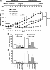
Synergistic antitumor effect of the combination of 5-Aza-CdR and castration in vivo. LNCaP cells were inoculated into the right thighs of BALB/c nude mice. When the tumor size reached to 250 mm3, the mice received an intraperitoneal injection of PBS (control), 5-Aza-CdR (0.25 mg/kg) thrice weekly, castration, or the combination of castration and 5-Aza-CdR (n = 12 per group). The tumor volume was monitored every two or three days (A). The animals were euthanized on days 14 and 35 post-treatment (n = 6 per group at either time point) for DNA, RNA, and protein analyses. The tumor weights were determined (B). PSA release into serum was measured by an ELISA analysis (C). The data are the mean ± SD of one representative experiment. Similar results were obtained in three independent experiments. *P < 0.05, **P < 0.01 compared to the control mice.
Figure 3
Figure 3
miR-146a expression in human prostate cancer xenograft mouse models. The miR-146a level was detected using qRT-PCR and described as the fold-change after normalization to U6 RNA in the tumor tissues of different mice groups (n = 6). The tumor tissue from the control mice of the first analysis was used to the calibrator samples. The data are the mean ± SD of one representative experiment. Similar results were obtained in three independent experiments. *P < 0.05, **P < 0.01 compared to the control samples.
Figure 4
Figure 4
The combination of 5-Aza-CdR and castration decreased the global gene methylation levels in human prostate cancer xenograft mouse models. (A) The expression of DNMT1, 3a and 3b was measured by qRT-PCR and normalized to the expression of GAPDH in each sample of mice (n = 6). (B) A schematic illustration of COBRA LINE-1. DNA was extracted from the xenograft tissues, treated with bisulfite, and subjected to PCR. LINE-1 methylation level was assessed by TasI-TaqI double digestion within the 160-bp amplicon. The methylated amplicons (TaqI positive) yielded two 80-bp DNA fragments, whereas the unmethylated amplicons (TasI positive) yielded 63- and 97-bp fragments. (C) COBRA LINE-1 of xenograft tissues. The percentage of methylation is listed above each test. The data are the mean ± SD of one representative experiment. Similar results were obtained in three independent experiments. *P < 0.05 and **P < 0.01 compared to the controls.
Figure 5
Figure 5
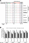
Comparing the methylation levels of four CpG sites located in the miR-146a promoter collected from samples from the different group by direct BSP sequencing. (A) Representative genomic sequencing chromatograms of the miR-146a promoter for each group. DNA was first treated with sodium bisulfite, and the amplified PCR products were then directly subjected to sequencing. The upper sequence is the untreated miR-146a promoter sequence; the lower sequences are treated sequences in different groups. The open boxes indicate the CpG sites. (B) Percentages of the methylation of four CpG sites in the miR-146a promoter. The methylation percentage of the individual CpG sites was calculated by the peak height of methylated residues (cytosine, C) divided by the sum of methylated and unmethylated residues (thymine, T). The methylation rate (%) is represented as the average ratio of methylated cytosines to total cytosines (methylated plus unmethylated) of all samples in each group. The data are the mean ± SD, n = 4–6 mice per group. *P < 0.05 and **P < 0.01 compared to the controls.

References

    1. Haas GP, Delongchamps N, Brawley OW, Wang CY, de la Roza G. The worldwide epidemiology of prostate cancer: perspectives from autopsy studies. Can J Urol. 2008;15(1):3866–3871. - PMC - PubMed
    1. Goldenberg SL, Gleave ME, Taylor D, Bruchovsky N. Clinical experience with intermittent androgen suppression in prostate cancer: minimum of 3 Years’ follow-Up. Mol Urol. 1999;3(3):287–292. - PubMed
    1. Jones PA, Baylin SB. The fundamental role of epigenetic events in cancer. Nat Rev Genet. 2002;3(6):415–428. - PubMed
    1. Haldrup C, Mundbjerg K, Vestergaard EM, Lamy P, Wild P, Schulz WA, Arsov C, Visakorpi T, Borre M, Hoyer S, Orntoft TF, Sørensen KD. DNA methylation signatures for prediction of biochemical recurrence after radical prostatectomy of clinically localized prostate cancer. J Clin Oncol. 2013;31(26):3250–3258. doi: 10.1200/JCO.2012.47.1847. - DOI - PubMed
    1. Patra SK, Bettuzzi S. Epigenetic DNA-(cytosine-5-carbon) modifications: 5-aza-2’-deoxycytidine and DNA-demethylation. Biochemistry Biokhimiia. 2009;74(6):613–619. doi: 10.1134/S0006297909060042. - DOI - PubMed

Publication types

LinkOut - more resources