. 2014 May 16:13:68.
doi: 10.1186/1475-2859-13-68.

A variant of green fluorescent protein exclusively deposited to active intracellular inclusion bodies

Affiliations

A variant of green fluorescent protein exclusively deposited to active intracellular inclusion bodies

Govindan Raghunathan et al. Microb Cell Fact. .

Abstract

Background: Inclusion bodies (IBs) were generally considered to be inactive protein deposits and did not hold any attractive values in biotechnological applications. Recently, some IBs of recombinant proteins were confirmed to show their functional properties such as enzyme activities, fluorescence, etc. Such biologically active IBs are not commonly formed, but they have great potentials in the fields of biocatalysis, material science and nanotechnology.

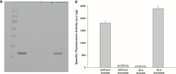
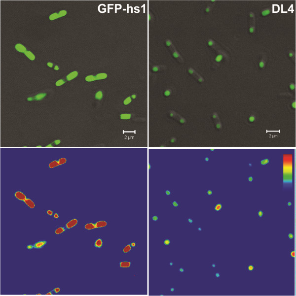
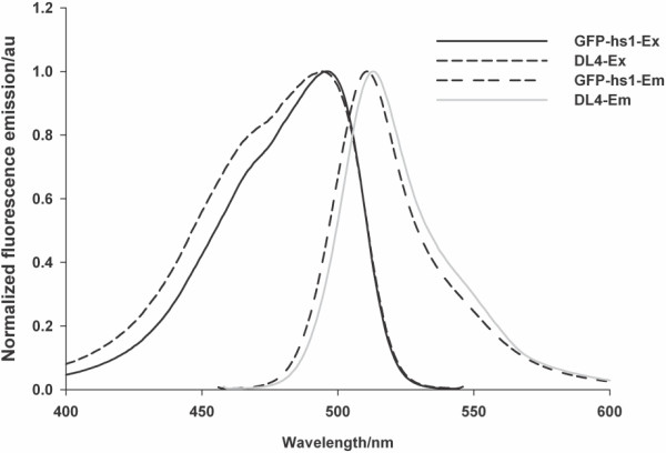
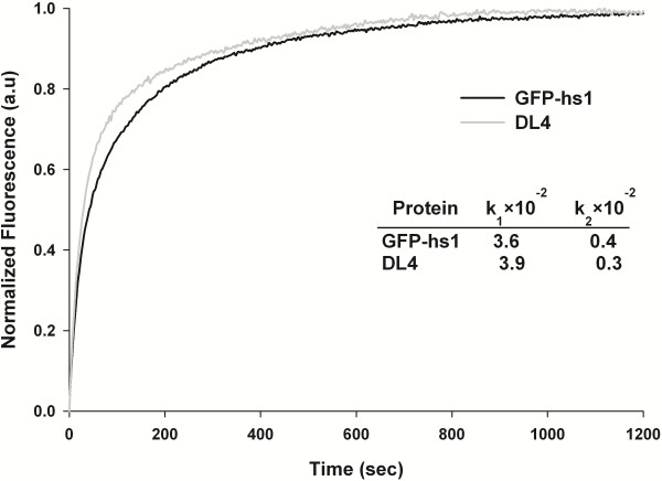
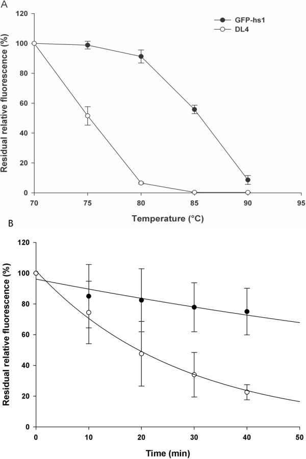
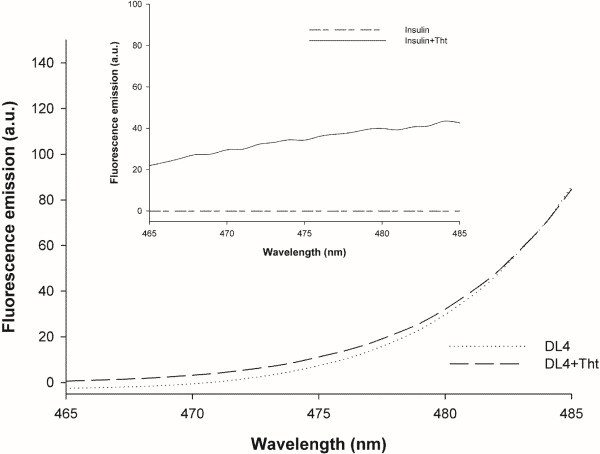
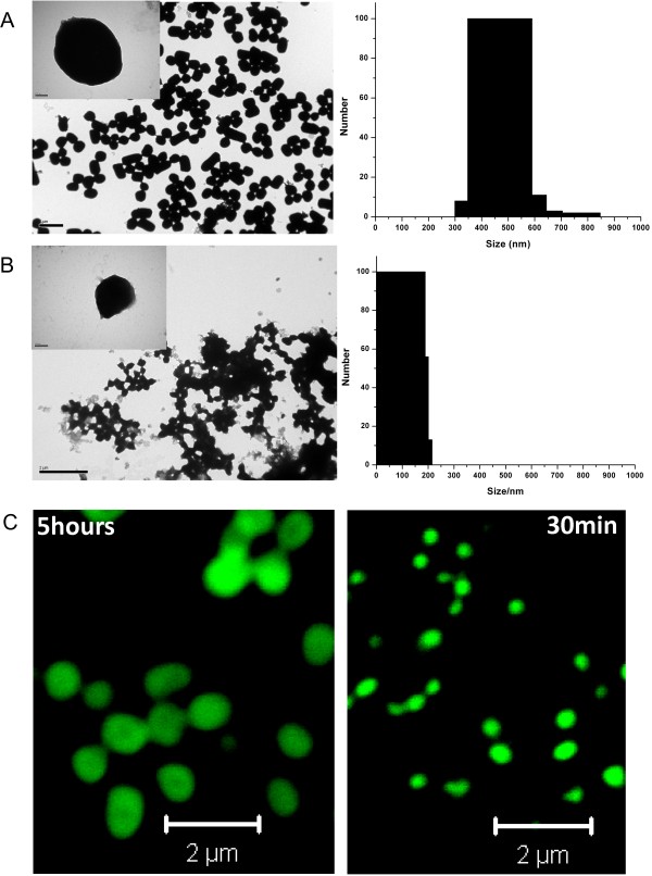
Results: In this study, we characterized the IBs of DL4, a deletion variant of green fluorescent protein which forms active intracellular aggregates. The DL4 proteins expressed in Escherichia coli were exclusively deposited to IBs, and the IBs were estimated to be mostly composed of active proteins. The spectral properties and quantum yield of the DL4 variant in the active IBs were almost same with those of its native protein. Refolding and stability studies revealed that the deletion mutation in DL4 didn't affect the folding efficiency of the protein, but destabilized its structure. Analyses specific for amyloid-like structures informed that the inner architecture of DL4 IBs might be amorphous rather than well-organized. The diameter of fluorescent DL4 IBs could be decreased up to 100-200 nm by reducing the expression time of the protein in vivo.

Conclusions: To our knowledge, DL4 is the first GFP variant that folds correctly but aggregates exclusively in vivo without any self-aggregating/assembling tags. The fluorescent DL4 IBs have potentials to be used as fluorescent biomaterials. This study also suggests that biologically active IBs can be achieved through engineering a target protein itself.


Figures

Figure 1
Figure 1
Expression and functional analysis of GFP-hs1 and DL4. (A) Soluble and insoluble expression profiles of GFP-hs1 and DL4 by SDS-PAGE. M – Marker, Lane 1,3 - soluble fractions of GFP-hs1 & DL4, Lane 2,4 – insoluble fractions of GFP-hs1 & DL4. (B) Specific fluorescence activities of DL4 and GFP-hs1 in their soluble and insoluble fractions. Specific fluorescence activity is defined as the fluorescence per microgram of protein. The values are the average of three measurements and standard deviations are shown.
Figure 2
Figure 2
In vivoanalysis of IBs formation. (Top) Analysis of protein distributions in E. coli using confocal microscope. Protein aggregates were observed as dense molecules forming localized pattern of distribution (DL4) whereas the expression of soluble protein was spread uniformly over the cytoplasm (GFP-hs1). Scale bar represents 2 μm. (Bottom) The green fluorescence intensity mapped in color scale from low (blue) to high (red).
Figure 3
Figure 3
Excitation/emission spectra of GFP-hs1 and DL4. Protein samples were scanned for excitation (between 400 & 550 nm) and emission spectra (between 450 & 600 nm) using fluorescence spectrophotometer.
Figure 4
Figure 4
Refolding kinetics of DL4 and GFP-hs1. The plot shows the refolding progression curve of DL4 and GFP-hs1, measured after complete denaturation in urea followed by renaturation initiated rapidly by dilution. Inset table shows the refolding rates of GFP variants. Normalized fluorescence in arbitrary units (au) was plotted against time in seconds.
Figure 5
Figure 5
Thermal stability of DL4 and GFP-hs1. (A) Effect of various temperatures on the stability of DL4 and GFP-hs1. The stability was measured by incubating at different temperatures for 30 minutes and the remaining fluorescence was plotted against temperature as function. (Error bar – Standard deviation of three experiments). (B) Time-dependent temperature effect on the stability of DL4 and GFP-hs1 at 80°C. The fluorescence at time zero min was taken as 100%. (Error bar – Standard deviation of two experiments).
Figure 6
Figure 6
Thioflavin T dye binding assay. Emission spectra of DL4 aggregates with and without ThT binding. The excitation wavelength was 450 nm. Inset shows the emission spectra of insulin aggregates with and without ThT binding as positive control.
Figure 7
Figure 7
Morphology of DL4 IBs before and after proteinase K treatment. (A) Electron micrograph of purified DL4 IBs (scale bar 2 μm) (B) Electron micrograph of DL4 IBs after proteinase K treatment. The scale bar depicts 1 μm.
Figure 8
Figure 8
Fluorescent protein nanoparticles based on DL4 IBs. Particle sizes and fluorescent images of purified IBs were analyzed by electron microscope, dynamic light scattering and confocal imaging. (A) Negative stained TEM image and DLS data of the purified DL4 expressed for 5 hours (B) Negative stained image and DLS data of purified DL4 expressed for 30 min. (Scale bar 2 μm). Inset figures show single IB of DL4 (scale bar 100 nm). (C) Confocal microscope images of the purified fluorescent DL4 particles expressed for 5 hours and 30 min (Scale bar 2 μm).

Similar articles

Cited by

References

    1. Villaverde A, Carrio MM. Protein aggregation in recombinant bacteria: biological role of inclusion bodies. Biotechnol Lett. 2003;25:1385–1395. doi: 10.1023/A:1025024104862. LA - English. - DOI - PubMed
    1. Fink AL. Protein aggregation: folding aggregates, inclusion bodies and amyloid. Fold Des. 1998;3:R9–R23. doi: 10.1016/S1359-0278(98)00002-9. - DOI - PubMed
    1. Baneyx F, Mujacic M. Recombinant protein folding and misfolding in Escherichia coli. Nat Biotechnol. 2004;22:1399–1408. doi: 10.1038/nbt1029. - DOI - PubMed
    1. Esposito D, Chatterjee DK. Enhancement of soluble protein expression through the use of fusion tags. Curr Opin Biotechnol. 2006;17:353–358. doi: 10.1016/j.copbio.2006.06.003. - DOI - PubMed
    1. Fink AL. Chaperone-mediated protein folding. Physiol Rev. 1999;79:425–449. - PubMed

Publication types

MeSH terms

Substances