. 2014 May 3:14:310.
doi: 10.1186/1471-2407-14-310.

Matrix metalloproteinase-10 promotes tumor progression through regulation of angiogenic and apoptotic pathways in cervical tumors

Affiliations

Matrix metalloproteinase-10 promotes tumor progression through regulation of angiogenic and apoptotic pathways in cervical tumors

Ge Zhang et al. BMC Cancer. .

Abstract

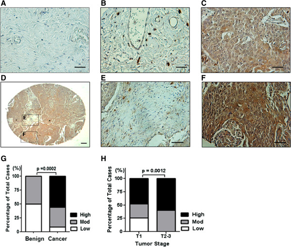
Background: Cancer invasion and metastasis develops through a series of steps that involve the loss of cell to cell and cell to matrix adhesion, degradation of extracellular matrix and induction of angiogenesis. Different protease systems (e.g., matrix metalloproteinases, MMPs) are involved in these steps. MMP-10, one of the lesser studied MMPs, is limited to epithelial cells and can facilitate tumor cell invasion by targeting collagen, elastin and laminin. Enhanced MMP-10 expression has been linked to poor clinical prognosis in some cancers, however, mechanisms underlying a role for MMP-10 in tumorigenesis and progression remain largely unknown. Here, we report that MMP-10 expression is positively correlated with the invasiveness of human cervical and bladder cancers.

Methods: Using commercial tissue microarray (TMA) of cervical and bladder tissues, MMP-10 immunohistochemical staining was performed. Furthermore using a panel of human cells (HeLa and UROtsa), in vitro and in vivo experiments were performed in which MMP-10 was overexpressed or silenced and we noted phenotypic and genotypic changes.

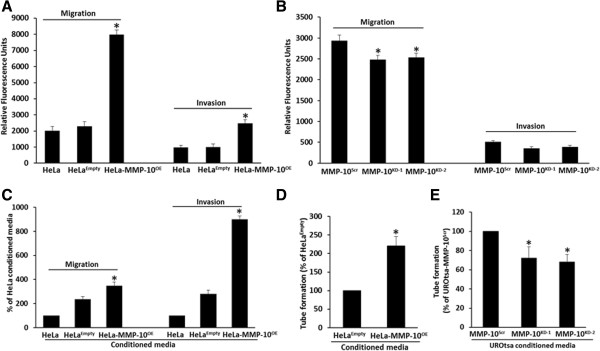
Results: Experimentally, we showed that MMP-10 can regulate tumor cell migration and invasion, and endothelial cell tube formation, and that MMP-10 effects are associated with a resistance to apoptosis. Further investigation revealed that increasing MMP-10 expression stimulates the expression of HIF-1α and MMP-2 (pro-angiogenic factors) and PAI-1 and CXCR2 (pro-metastatic factors), and accordingly, targeting MMP-10 with siRNA in vivo resulted in diminution of xenograft tumor growth with a concomitant reduction of angiogenesis and a stimulation of apoptosis.

Conclusion: Taken together, our findings show that MMP-10 can play a significant role in tumor growth and progression, and that MMP-10 perturbation may represent a rational strategy for cancer treatment.


Figures

Figure 1
Figure 1
MMP-10 expression is high in cervical cancer tissue. Representative images of benign cervical tissue (A), cervical tumor with weak (B), and cervical tumor with strong (C) MMP-10 expression (brown). Representative image of a cervical tumor core on a tissue microarray (D), MMP-10 expression in the tumor-associated stroma (E), and in the tumor epithelia (F). Scale bars, 100 μm. G, quantification of MMP-10 expression levels in benign cervical tissue (n = 10) vs. cervical tumor (n = 70). Increased MMP-10 expression was noted in cervical tumors compared to benign cervical tissue. H, quantification of MMP-10 expression levels in low stage cervical tumors vs. high stage cervical tumors. Increased MMP-10 expression was noted in high stage cervical tumors.
Figure 2
Figure 2
MMP-10 expression in HeLa and UROtsa human cell lines. A, MMP-10 expression was assessed by quantitative reverse transcriptase-PCR (qPCR), and immunoblot (IB) analyses in HeLa and UROtsa cells. MMP-10 activity was assessed by zymography, demonstrated by the degradation of casein. B, MMP-10 expression and activity was assessed by qPCR, immunoblot analyses and zymogram in stable HeLa clones; HeLa-MMP-10OE overexpressing a functionally human MMP-10 in a pCMV6-Entry vector, and HeLaEmpty an empty-vector transfected control. C, MMP-10 assessed by qPCR, immunoblot and zymography in UROtsa clones; UROtsa-MMP-10KD-1, UROtsa-MMP-10KD-2 functional knockdown clones transfected with MMP-10 siRNA, and UROtsa-MMP-10Scr transfected with a scrambled siRNA control.
Figure 3
Figure 3
MMP-10 promotes cellular migration and invasion. A, HeLa cells (parental, HeLaEmpty and HeLa-MMP-10OE) and B, UROtsa cells (parental, UROtsa-MMP-10KD-1, UROtsa-MMP-10KD-2 and UROtsa-MMP-10Scr) were subjected to in vitro migration and invasion assays. Data are average values and SDs of three independent experiments conducted in triplicate. Overexpression of MMP-10 in HeLa-MMP-10OE cells was noted to correspond with an increase in migratory and invasive potential, while silencing of MMP-10 in UROtsa-MMP-10KD-1 resulted in a reduced migratory potential. C, HUVEC cells were placed in the invasion chamber and then exposed to conditioned media in the lower chamber from HeLa cells (parental, HeLa-MMP-10OE and HeLaEmpty), and UROtsa cells (UROtsa-MMP-10KD-1, UROtsa-MMP-10KD-2 and UROtsa-MMP-10Scr) for in vitro migration and invasion assays. Data are average values and SDs of three independent experiments conducted in triplicate. Conditioned media from HeLa-MMP-10OE cells that overexpressed MMP-10 was noted to induce migration and invasion of HUVEC cells. D, Capillary tube formation of HUVECs cultured in conditioned media from HeLa-MMP-10OE and HeLaEmpty cells were quantified as total tube length per field in micrometers and was noted to be enhanced when MMP-10 was overexpressed. Data are average values and SDs of three independent experiments conducted in triplicate. E, Capillary tube formation in HUVECs cultured in conditioned media from UROtsa-MMP-10KD-1, UROtsa-MMP-10KD-2 and UROtsa-MMP-10Scr cells were quantified as total tube length per field in micrometers and was noted to be inhibited when MMP-10 was silenced. Data are average values and SDs of three independent experiments conducted in triplicate. *, p < 0.05.
Figure 4
Figure 4
MMP-10 regulates the expression of key angiogenic and metastatic factors. A, Angiogenesis RT2 Profiler PCR Arrays evaluated by quantitative RT-PCR assessed the expression of 84 gene transcripts. Gene expression levels were normalized to the combination of five housekeeping genes (β-actin, B2M, GAPDH, HPRT and RPLP). Changes in expression of HIF-1α, MMP-9, MMP-2, IGF-1 transcripts (see other changes listed in Additional file 3: Table S1) were noted in HeLa-MMP-10OE cells relative to HeLaEmpty cells. B, Western blot analysis confirmed the differential expression of HIF-1α and MMP-9 at the protein level. GAPDH served as a loading control. C, Metastasis RT2 Profiler PCR Arrays evaluated by quantitative RT-PCR assessed 84 gene transcripts. Gene expression levels were normalized to the combination of five housekeeping genes (β-actin, B2M, GAPDH, HPRT and RPLP). Changes in expression of Src, MMP-2, PAI-1, CXCR2 (also see Additional file 4: Table S2) were noted in HeLa-MMP-10OE cells relative to HeLaEmpty cells. D, Western blot analysis confirmed differential protein expression of PAI-1 and CXCR2. GAPDH served as a loading control.
Figure 5
Figure 5
Overexpression of MMP-10 results in inhibition of apoptosis. A, Caspase activity assay was performed with HeLa-MMP-10OE and HeLaEmpty cells after exposing the cells to the apoptotic-inducing agent, staurosporine at 1 μM. At least three independent experiments performed in triplicate were used to calculate mean ± SD values. *, p < 0.05. The apoptotic effects of staurosporine were abrogated by MMP-10 overexpression. B, Annexin V apoptotic assays were also performed with HeLa-MMP-10OE and HeLaEmpty cells as described above. Assays confirmed that high expression of MMP-10 was associated with resistance to induced apoptosis. C, Changes in mitochondrial membrane potential (DCm) in HeLa-MMP-10OE and control HeLaEmpty cells were assessed by flow cytometric analysis. At least three independent experiments performed in triplicate wells were used to calculate mean ± SD values. *, p < 0.05. High expression of MMP-10 was associated with a significant increase in DCm compared to control. D, Western blot analysis was performed on the above cell lines confirming MMP-10 overexpression was associated with the expression of apoptosis factors. For example, the expression of key intrinsic apoptotic pathway factors (Bcl-xl, Bax and Bak), and factors in the extrinsic apoptotic pathway (FasL and cleaved caspase-8) were altered when MMP-10 was overexpressed. GAPDH served as controls. E, The expression of the proteins from the Western blot from panel D was quantitated and plotted to illustrate relative expression of the apoptotic associated proteins.
Figure 6
Figure 6
MMP-10 regulates HeLa xenograft tumorigenicity. A, Human and mouse MMP-10 expression in subcutaneous HeLa tumors and in parental HeLa cells grown in culture was assessed by qPCR. Gene expression level was normalized to GAPDH. Cultured parental HeLa cells expressed minimal MMP-10, but when grown in a subcutaneous mouse model, increases in both human and mouse MMP-10 were noted. B, Tumor growth was established by subcutaneous injection of parental HeLa cells into athymic nude mice (nu/nu). PBS (negative control), scrambled siRNA (negative control), human MMP-10 siRNA and murine MMP-10 siRNA reagents were injected intratumorally twice weekly as described in Materials and Methods. Tumor size was plotted as mean ± SD from the four treatment groups (n = 10/group). Treatment with siRNA MMP-10 human alone, siRNA MMP-10 mouse alone and the combination of human and mouse siRNA MMP-10 were associated with a significant reduction in tumor burden. *, p < 0.05. C, Human MMP-10 and mouse MMP-10 expression in excised tumors are shown (images 400 ×, Bar = 50 μm). A reduction in MMP-10 and CD31 expression along with an induction of cleaved caspase-3 was noted in tumors treated with human MMP-10 siRNA alone, mouse MMP-10 siRNA alone and the combination of human and mouse siRNAs. D, MMP-10 expression was quantified based on MMP-10 staining. E, Microvessel density (MVD) was quantified based on CD31 staining. Apoptotic index (AI) was quantified based on cleaved caspase-3 staining. Data are presented as mean ± SD, *, p < 0.05 compared to negative control.

Similar articles

Cited by

References

    1. Hojilla CV, Mohammed FF, Khokha R. Matrix metalloproteinases and their tissue inhibitors direct cell fate during cancer development. Br J Canc. 2003;89:1817–1821. doi: 10.1038/sj.bjc.6601327. - DOI - PMC - PubMed
    1. Egeblad M, Werb Z. New functions for the matrix metalloproteinases in cancer progression. Nat Rev Cancer. 2002;2:161–174. doi: 10.1038/nrc745. - DOI - PubMed
    1. Madlener M, Mauch C, Conca W, Brauchle M, Parks WC, Werner S. Regulation of the expression of stromelysin-2 by growth factors in keratinocytes: implications for normal and impaired wound healing. Biochem J. 1996;320:659–664. - PMC - PubMed
    1. Chakraborti S, Mandal M, Das S, Mandal A, Chakraborti T. Regulation of matrix metalloproteinases: an overview. Mol Cell Biochem. 2003;253:269–285. doi: 10.1023/A:1026028303196. - DOI - PubMed
    1. Krampert M, Bloch W, Sasaki T, Bugnon P, Rülicke T, Wolf E, Bloch W, Sasaki T, Bugnon P, Rülicke T, Wolf E. Activities of the matrix metalloproteinase stromelysin-2 (MMP-10) in matrix degradation and keratinocyte organization in wounded skin. Mol Biol Cell. 2004;15:5242–5254. doi: 10.1091/mbc.E04-02-0109. - DOI - PMC - PubMed

Publication types

MeSH terms

Substances