

Retinal nerve fibre layer, ganglion cell layer and choroid thinning in migraine with aura
- PMID: 24885597
- PMCID: PMC4229806
- DOI: 10.1186/1471-2415-14-75
Retinal nerve fibre layer, ganglion cell layer and choroid thinning in migraine with aura
Abstract
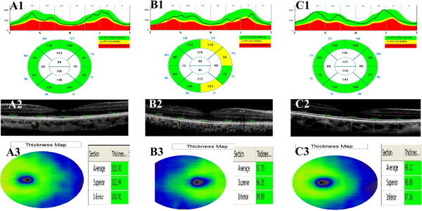
Background: The aim of this study was to investigate the thickness of the retinal nerve fiber layer (RNFL), the ganglion cell layer (GCL), and choroid thickness (CT) in patients who have migraines, with and without aura, using spectral optical coherence tomography (OCT).
Methods: Forty-five patients who had migraines without aura (Group 1), 45 patients who had migraines with aura (Group 2), and 30 healthy participants (control group) were included in the study. Spectral OCT was used to measure the RNFL, GCL and CT values for all patients.
Results: The mean age of Group 1, Group 2, and the control group was 34.6 ± 4.3, 32.8 ± 4.9, and 31.8 ± 4.6 years, respectively. The mean attack frequency was 3.6/month in Group 1 and 3.7/month in Group 2. The mean age among the groups (p = 0.27) and number of attacks in migraine patients (p = 0.73) were not significantly different. There was significant thinning in the RNFL and GCL in Group 2 (p < 0.05, p < 0.001 respectively), while there were no significant differences in RNFL and GCL measurements between Group 1 and the control group (p > 0.05). All groups were significantly different from one another with respect to CT, with the most thinning observed in Group 2 (p < 0.001). When all migraine patients (without grouping) were compared with the control group, there were significant differences on all parameters: RNFL thickness, GCC thickness and CT (p < 0.05).
Conclusions: RNFL and GCL were significantly thinner in the migraine patients with aura as compared with both the migraine patients without aura and the control subjects. In migraine, both with aura and without aura, patients' choroid thinning should be considered when evaluating ophthalmological findings.
Figures
Similar articles
-
Evaluation of retinal nerve fibre layer, ganglion cell layer and choroidal thickness with optical coherence tomography in migraine patients: a case-control study.Clin Exp Optom. 2018 Jan;101(1):109-115. doi: 10.1111/cxo.12585. Epub 2017 Sep 20. Clin Exp Optom. 2018. PMID: 28940251
-
Retinal nerve fiber layer, ganglion cell complex, and choroidal thicknesses in migraine.Arq Bras Oftalmol. 2016 Apr;79(2):78-81. doi: 10.5935/0004-2749.20160024. Arq Bras Oftalmol. 2016. PMID: 27224067
-
Neuroretinal evaluation using optical coherence tomography in patients affected by pituitary tumors.Ann Ital Chir. 2017;88:7-14. Ann Ital Chir. 2017. PMID: 28447589
-
Evaluation of retinal nerve fiber layer, ganglion cell layer and macular changes in patients with migraine.Acta Neurol Belg. 2017 Mar;117(1):121-129. doi: 10.1007/s13760-016-0715-1. Epub 2016 Oct 21. Acta Neurol Belg. 2017. PMID: 27770392
-
Macula and retinal nerve fiber layer in migraine patients: analysis by spectral domain optic coherence tomography.Semin Ophthalmol. 2015 Mar;30(2):124-8. doi: 10.3109/08820538.2013.833270. Epub 2013 Oct 30. Semin Ophthalmol. 2015. PMID: 24171810
Cited by
-
Evaluation of the lamina cribrosa thickness and depth in patients with migraine.Int Ophthalmol. 2020 Jan;40(1):89-98. doi: 10.1007/s10792-019-01160-2. Epub 2019 Aug 20. Int Ophthalmol. 2020. PMID: 31432353
-
Comment on Retinal findings of COVID-19 patients using ocular coherence tomography angiography two to three months after infection: Ocular appearance recovered COVID-19 patient.Photodiagnosis Photodyn Ther. 2022 Sep;39:102908. doi: 10.1016/j.pdpdt.2022.102908. Epub 2022 May 14. Photodiagnosis Photodyn Ther. 2022. PMID: 35577061 Free PMC article. No abstract available.
-
Is Retinal Nerve Fiber Layer Thickness Change Related to Headache Lateralization in Migraine?Korean J Ophthalmol. 2016 Apr;30(2):134-9. doi: 10.3341/kjo.2016.30.2.134. Epub 2016 Mar 25. Korean J Ophthalmol. 2016. PMID: 27051262 Free PMC article.
-
Foveal and Peripapillary Vascular Decrement in Migraine With Aura Demonstrated by Optical Coherence Tomography Angiography.Invest Ophthalmol Vis Sci. 2017 Oct 1;58(12):5477-5484. doi: 10.1167/iovs.17-22477. Invest Ophthalmol Vis Sci. 2017. PMID: 29059314 Free PMC article.
-
Evaluation of Retinal and Posterior Segment Vascular Changes Due to Systemic Hypoxia Using Optical Coherence Tomography Angiography.J Clin Med. 2024 Nov 7;13(22):6680. doi: 10.3390/jcm13226680. J Clin Med. 2024. PMID: 39597827 Free PMC article.
References
-
- Tan FU, Akarsu C, Güllü R. Retinal nerve fiber layer thickness is unaffected in migraine patients. Acta Neurol Scand. 2005;112:19–23. - PubMed
-
- Totan Y, Çekiç O. Görme ile ilgili elektrofizyolojik testler ve klinik uygulamaları. (Vision-related Electrophysiological tests and their clinical applications. MN Oftalmol. 1996;3:195–198.
Publication types
MeSH terms
LinkOut - more resources
Full Text Sources
Other Literature Sources
Miscellaneous
