. 2014 May 30:12:44.
doi: 10.1186/1741-7007-12-44.

Molecular dissection of Wnt3a-Frizzled8 interaction reveals essential and modulatory determinants of Wnt signaling activity

Affiliations

Molecular dissection of Wnt3a-Frizzled8 interaction reveals essential and modulatory determinants of Wnt signaling activity

Sumit Kumar et al. BMC Biol. .

Abstract

Background: Wnt proteins are a family of secreted signaling molecules that regulate key developmental processes in metazoans. The molecular basis of Wnt binding to Frizzled and LRP5/6 co-receptors has long been unknown due to the lack of structural data on Wnt ligands. Only recently, the crystal structure of the Wnt8-Frizzled8-cysteine-rich-domain (CRD) complex was solved, but the significance of interaction sites that influence Wnt signaling has not been assessed.

Results: Here, we present an extensive structure-function analysis of mouse Wnt3a in vitro and in vivo. We provide evidence for the essential role of serine 209, glycine 210 (site 1) and tryptophan 333 (site 2) in Fz binding. Importantly, we discovered that valine 337 in the site 2 binding loop is critical for signaling without contributing to binding. Mutations in the presumptive second CRD binding site (site 3) partly abolished Wnt binding. Intriguingly, most site 3 mutations increased Wnt signaling, probably by inhibiting Wnt-CRD oligomerization. In accordance, increasing amounts of soluble Frizzled8-CRD protein modulated Wnt3a signaling in a biphasic manner.

Conclusions: We propose a concentration-dependent switch in Wnt-CRD complex formation from an inactive aggregation state to an activated high mobility state as a possible modulatory mechanism in Wnt signaling gradients.


Figures

Figure 1
Figure 1
Structural model for mouse Wnt3a-Fz8 interaction. (A) Overview of the mouse Wnt3a-Fz8-CRD complex. Wnt3a is in grey, Fz8-CRD in blue. The binding sites (site 1, yellow; site 2, red; pseudo site 3, pink) are highlighted and the respective residues marked. Close-up views of binding sites 1 (B) and 2 (C), and pseudo site 3 (D). (E) Alignment of Xenopus Wnt8 and mouse Wnt3a sequences. Mutations introduced into site 1 (yellow), site 2 (red) and site 3 (pink) are indicated.
Figure 2
Figure 2
Effect of Wnt3a point mutants on signaling activity. (A, C, E) Wnt3a point mutants from sites 1, 2 and 3 were tested in the Wnt reporter assay for their activity and compared with wild-type Wnt3a. HEK293T cells were transiently transfected with luciferase-based TCF/Wnt reporter with wild-type Wnt3a or the indicated mutant constructs. (B, D, F) Secretion levels of Wnt3a mutants analyzed by Western blot. HEK293T cells were transfected with wild-type Wnt3a or the indicated mutant constructs. Supernatants and cell lysates were analyzed 48 hr after transfection. Membranes were probed with Wnt3a and α-tubulin (loading control) antibodies. (G) Effect of double mutants on signaling activity. (H) Secretion levels of Wnt3a double mutants in HEK293T cells. Experiments were carried out in triplicate. Error bars depict standard deviation. Statistical significance in relative luciferase activity levels (A, C, E, G) of Wnt3a mutants compared with wild-type Wnt3a is indicated in the following way: *P < 0.05, **P < 0.01, ***P < 0.001 and n.s. as not significant according to Student’s t-test.
Figure 3
Figure 3
Binding of Wnt3a mutants to Fz8-CRD-Fc. (A) Binding efficiency of mutants from sites 1, 2 and 3, and double mutants were analyzed by precipitation from a conditioned medium with recombinant Fz8-CRD-Fc or Fc alone pre-bound to Protein G agarose beads. Bound protein fractions were analyzed by immunoblotting using anti-Wnt3a antibody. (B) Pull-down assay of wild-types Wnt3a and site 1 mutants S209A and G210R with Blue Sepharose beads. BS, Blue Sepharose;s IgG, immunoglobulin G; IP, immunoprecipitation; CM, Conditioned Medium.
Figure 4
Figure 4
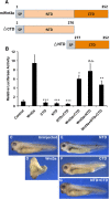
In vitro and in vivo activity of Wnt3a NTD and CTD constructs. (A) Schematic representation of applied mouse Wnt3a NTD and CTD constructs. (B) Wnt reporter assay in HEK293T showing the effect of the Wnt3a NTD and CTD on signaling activity. Experiments were carried out in triplicate. Error bars depict standard deviation. Statistical significance in relative luciferase activity levels compared to wild-type Wnt3a levels as indicated: *P < 0.05, **P < 0.01, ***P < 0.001 and n.s. as not significant according to Student’s t-test. (C-G)Xenopus embryos at 4-cell stage were injected into the ventral-marginal-zone with 3pg mRNA encoding wild type Wnt3a (D), NTD (E), CTD (F) or NTD + CTD (G).
Figure 5
Figure 5
Expression of mouse Wnt3a mutants in zebrafish embryos. (A) Lateral view of zebrafish embryos, 1.5 days post fertilization (dpf), upon injection of capped mRNA encoding wild-type and mutant mouse Wnt3a capped mRNAs. Note lack of forebrain, eye field and midbrain structures in lateral view. (B) Posteriorizing effect of Wnt3a overexpression on zebrafish development as analyzed at 1.5 dpf. Pictures are single optical sections either of lateral (upper panel) or medial (lower panel) sagittal sections, with staining for F-actin marking cell outlines and thereby general nervous system morphology. The weaker phenotype with loss of retina and forebrain, though remnants of midbrain structures might be present, is termed phenotype type I. The stronger phenotype (lacking the forebrain, retina, lens and midbrain) is phenotype type II. (C) Penetrance of the dorsalized phenotype in zebrafish embryogenesis on wild-type and mutant mouse Wnt3a expression, quantified as a percentage. Type I (weaker) and type II (stronger) respond to the phenotypes shown in Figure 5B. Total number of embryos (n) from three independent experiments: non-injected: 403, EF1a: 76, wild-type mouse Wnt3a: 224, S209A: 113, G210R: 96, F331A: 79, W333A: 54, C335A: 91, V337R: 139, V60R: 86, E68A: 64, A96R: 124, F169A: 82, E68A/F169A: 88 and F331A/W333A: 82. In all panels, anterior is to the left and dorsal is up. Eyes are outlined with dashed pink lines. fb, forebrain; hb, hindbrain; l, lens; mb, midbrain; ov, otic vesicle; r, retina; WT, wild type; mhb, midbrain-hindbrain boundary.
Figure 6
Figure 6
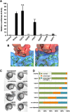
Influence of different V337 mutations on Wnt activity in vitro and in vivo. (A) Wnt reporter assay showing the influence of different amino acid side chains at position V337 on the signaling activity of Wnt3a. Position V337 is highly sensitive towards bulky side chains as illustrated in (B), which is an interaction model for the V337W mutant compared to the wild type. (C) Posteriorizing effect and (D) penetrance of the dorsalized phenotype in zebrafish embryogenesis upon expression of wild-type mouse Wnt3a and diverse V337 mutants. Experiments were carried out in triplicate. Error bars depict standard deviation. Statistical significance in relative luciferase activity levels compared to the wild-type Wnt3a levels in (A) as indicated: *P < 0.05, **P < 0.01, ***P < 0.001 and n.s. as not significant according to Student’s t-test. wt, wild type.
Figure 7
Figure 7
Modulation of Wnt signaling by soluble Fz8-CRD-Fc. (A) Wnt reporter assay showing the influence of purified mFz8-CRD-Fc on Wnt signaling in a dose-dependent manner. (B) Immunoblot depicting the influence of recombinant Fz8-CRD-Fc on the concentration of soluble Wnt3a protein in the cell supernatant. The non-binding Wnt3a mutant W333A was used as a control. For all conditions, cells were seeded at equal cell density. (C) Double luciferase chamber assay using CMV-β-galactosidase and Super TOPFlash reporter in HEK293 cells. The Fz8-CRD-Fc protein was applied at increasing concentrations to the Wnt-producing cells. Luciferase activity was monitored in Wnt3a responder cells. Inset shows the set-up of the chamber assay. Experiments were carried out in triplicate. Error bars depict standard deviation. Statistical significance in relative luciferase activity levels compared to the wild-type Wnt3a levels (A, C) as indicated: *P < 0.05, **P < 0.01, ***P < 0.001 and n.s. as not significant according to Student’s t-test. CRD, cysteine-rich domain; WB, Western blot.
Figure 8
Figure 8
Model for Wnt-CRD complex formation. (A) Sequential binding of membrane-associated Wnt ligands to CRDs. The first binding step is realized between the membrane-distal Wnt CTD and Fz-CRD or other CRD-related proteins via a site 2 interaction. This facilitates membrane detachment of the Wnt NTD and a switch of the lipid chain into the site 1 binding pocket. (B) Biphasic modulation of Wnt activity by CRD proteins. At low concentrations of CRD proteins, active Wnt-CRD complexes are formed by Wnt mobilization and transfer to Fz receptors. At high CRD concentrations, oligomerization is induced via a site 3 interaction, leading to trapping of Wnt ligands in inactive Wnt-CRD aggregates. CRD, cysteine-rich domain; CTD, C-terminal domain; NTD, N-terminal domain.

References

    1. Logan CY, Nusse R. The Wnt signaling pathway in development and disease. Annu Rev Cell Dev Biol. 2004;12:781–810. doi: 10.1146/annurev.cellbio.20.010403.113126. - DOI - PubMed
    1. MacDonald BT, Tamai K, He X. Wnt/beta-catenin signaling: components, mechanisms, and diseases. Dev Cell. 2009;12:9–26. doi: 10.1016/j.devcel.2009.06.016. - DOI - PMC - PubMed
    1. Holstein TW, Watanabe H, Ozbek S. Signaling pathways and axis formation in the lower metazoa. Curr Top Dev Biol. 2011;12:137–177. - PubMed
    1. Willert K, Brown JD, Danenberg E, Duncan AW, Weissman IL, Reya T, Yates JR 3rd, Nusse R. Wnt proteins are lipid-modified and can act as stem cell growth factors. Nature. 2003;12:448–452. doi: 10.1038/nature01611. - DOI - PubMed
    1. Port F, Kuster M, Herr P, Furger E, Banziger C, Hausmann G, Basler K. Wingless secretion promotes and requires retromer-dependent cycling of Wntless. Nat Cell Biol. 2008;12:178–185. doi: 10.1038/ncb1687. - DOI - PubMed

Publication types

MeSH terms