

A case of multiple hepatic angiomyolipomas with high (18) F-fluorodeoxyglucose uptake
- PMID: 24885757
- PMCID: PMC4036299
- DOI: 10.1186/1471-2342-14-17
A case of multiple hepatic angiomyolipomas with high (18) F-fluorodeoxyglucose uptake
Abstract
Background: Hepatic angiomyolipoma is a rare benign mesenchymal tumor. We report an unusual case of a patient with multiple hepatic angiomyolipomas exhibiting high (18) F-fluorodeoxyglucose (FDG) uptake.
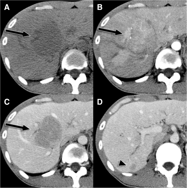
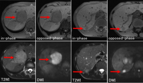
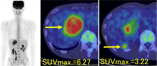
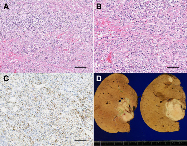
Case presentation: A 29-year-old man with a medical history of tuberous sclerosis was admitted to our hospital for fever, vomiting, and weight loss. Abdominal dynamic computed tomography revealed faint hypervascular hepatic tumors in segments 5 (67 mm) and 6 (10 mm), with rapid washout and clear borders; however, the tumors exhibited no definite fatty density. Abdominal magnetic resonance imaging revealed that the hepatic lesions were slightly hypointense on T1-weighted imaging, slightly hyperintense on T2-weighted imaging, and hyperintense with no apparent fat component on diffusion-weighted imaging. FDG-positron emission tomography (PET) imaging revealed high maximum standardized uptake values (SUVmax) of 6.27 (Segment 5) and 3.22 (Segment 6) in the hepatic tumors. A right hepatic lobectomy was performed, and part of the middle hepatic vein was also excised. Histological examination revealed that these tumors were characterized by the background infiltration of numerous inflammatory cells, including spindle-shaped cells, and a resemblance to an inflammatory pseudotumor. Immunohistochemical evaluation revealed that the tumor stained positively for human melanoma black-45. The tumor was therefore considered an inflammatory pseudotumor-like angiomyolipoma. Although several case reports of hepatic angiomyolipoma have been described or reviewed in the literature, only 3 have exhibited high (18) F-FDG uptake on PET imaging with SUVmax ranging from 3.3-4.0. In this case, increased (18) F-FDG uptake is more likely to appear, particularly if the inflammation is predominant.
Conclusion: Although literature regarding the role of (18) F-FDG-PET in hepatic angiomyolipoma diagnosis is limited and the diagnostic value of (18) F-FDG-PET has not yet been clearly defined, the possibility that hepatic angiomyolipoma might exhibit high (18) F-FDG uptake should be considered.
Figures
Similar articles
-
Hepatic epithelioid angiomyolipoma mimicking hepatocellular carcinoma on MR and 18F-FDG PET/CT imaging: A case report and literature review.Hell J Nucl Med. 2022 May-Aug;25(2):205-209. doi: 10.1967/s002449912480. Epub 2022 Aug 3. Hell J Nucl Med. 2022. PMID: 35913867 Review.
-
F-18 FDG PET/CT findings in two patients with hepatic angiomyolipoma with and without intratumoral hemorrhage.Clin Nucl Med. 2010 Jan;35(1):18-21. doi: 10.1097/RLU.0b013e3181c3611f. Clin Nucl Med. 2010. PMID: 20026966
-
High 18F-Fluorodeoxyglucose Uptake in Adrenal Angiomyolipoma: Case Report and Review of Literature.Medicine (Baltimore). 2015 Jun;94(22):e900. doi: 10.1097/MD.0000000000000900. Medicine (Baltimore). 2015. PMID: 26039121 Free PMC article. Review.
-
More advantages in detecting bone and soft tissue metastases from prostate cancer using 18F-PSMA PET/CT.Hell J Nucl Med. 2019 Jan-Apr;22(1):6-9. doi: 10.1967/s002449910952. Epub 2019 Mar 7. Hell J Nucl Med. 2019. PMID: 30843003
-
Hepatic Epithelioid Angiomyolipoma and 18F-FDG PET/CT.Clin Nucl Med. 2018 Jun;43(6):422-424. doi: 10.1097/RLU.0000000000002048. Clin Nucl Med. 2018. PMID: 29578870
Cited by
-
Clinical characteristics and outcomes of patients with hepatic angiomyolipoma: A literature review.World J Gastroenterol. 2021 May 21;27(19):2299-2311. doi: 10.3748/wjg.v27.i19.2299. World J Gastroenterol. 2021. PMID: 34040323 Free PMC article. Review.
-
18F-Fluorodeoxyglucose Positron Emission Tomography-Computed Tomography in Response Assessment of Perivascular Epithelioid Cell Tumor of the Pelvic Cavity to Irinotecan and Temozolomide.Indian J Nucl Med. 2020 Oct-Dec;35(4):348-349. doi: 10.4103/ijnm.IJNM_29_20. Epub 2020 Oct 21. Indian J Nucl Med. 2020. PMID: 33642765 Free PMC article.
-
The role of (18)F-FDG PET/CT imaging in patient with malignant PEComa treated with mTOR inhibitor.Onco Targets Ther. 2015 Jul 30;8:1967-70. doi: 10.2147/OTT.S85444. eCollection 2015. Onco Targets Ther. 2015. PMID: 26257526 Free PMC article.
-
Unresectable hepatic PEComa: a rare malignancy treated with stereotactic body radiation therapy (SBRT) followed by complete resection.Radiat Oncol. 2018 Feb 20;13(1):28. doi: 10.1186/s13014-018-0974-5. Radiat Oncol. 2018. PMID: 29463266 Free PMC article.
-
Characteristics and Treatment of Primary Hepatic Perivascular Epithelioid Cell Tumor (PEComa) in Adults: A Systematic Review.Cancers (Basel). 2025 Jul 8;17(14):2276. doi: 10.3390/cancers17142276. Cancers (Basel). 2025. PMID: 40723161 Free PMC article. Review.
References
-
- Butte JM, Do RK, Shia J, Gönen M, D’Angelica MI, Getrajdman GI, Allen PJ, Fong Y, Dematteo RP, Klimstra DS, Jarnagin WR. Liver angiomyolipomas: a clinical, radiologic, and pathologic analysis of 22 patients from a single center. Surgery. 2011;150:557–67. doi: 10.1016/j.surg.2011.03.006. - DOI - PubMed
-
- Blasiak B, Barnes S, Foniok T, Rushforth D, Matyas J, Ponjevic D, Weglarz WP, Tyson R, Iqbal U, Abulrob A, Sutherland GR, Obenaus A, Tomanek B. Comparison of T2 and T2*-weighted MR molecular imaging of a mouse model of glioma. BMC Med Imaging. 2013;13:20. doi: 10.1186/1471-2342-13-20. - DOI - PMC - PubMed
-
- Jiang T-A, Zhao Q-Y, Chen M-Y, Wang L-J, Ao J-Y. Diagnostic analysis of hepatic angiomyolipoma. Hepatobiliary Pancreat Dis Int. 2005;4:152–5. - PubMed
MeSH terms
Substances
LinkOut - more resources
Full Text Sources
Other Literature Sources
Medical
