. 2014 May 16:15:8.
doi: 10.1186/1471-2199-15-8.

Differential regulation of the α-globin locus by Krüppel-like Factor 3 in erythroid and non-erythroid cells

Affiliations

Differential regulation of the α-globin locus by Krüppel-like Factor 3 in erythroid and non-erythroid cells

Alister P W Funnell et al. BMC Mol Biol. .

Abstract

Background: Krüppel-like Factor 3 (KLF3) is a broadly expressed zinc-finger transcriptional repressor with diverse biological roles. During erythropoiesis, KLF3 acts as a feedback repressor of a set of genes that are activated by Krüppel-like Factor 1 (KLF1). Noting that KLF1 binds α-globin gene regulatory sequences during erythroid maturation, we sought to determine whether KLF3 also interacts with the α-globin locus to regulate transcription.

Results: We found that expression of a human transgenic α-globin reporter gene is markedly up-regulated in fetal and adult erythroid cells of Klf3-/- mice. Inspection of the mouse and human α-globin promoters revealed a number of canonical KLF-binding sites, and indeed, KLF3 was shown to bind to these regions both in vitro and in vivo. Despite these observations, we did not detect an increase in endogenous murine α-globin expression in Klf3-/- erythroid tissue. However, examination of murine embryonic fibroblasts lacking KLF3 revealed significant de-repression of α-globin gene expression. This suggests that KLF3 may contribute to the silencing of the α-globin locus in non-erythroid tissue. Moreover, ChIP-Seq analysis of murine fibroblasts demonstrated that across the locus, KLF3 does not occupy the promoter regions of the α-globin genes in these cells, but rather, binds to upstream, DNase hypersensitive regulatory regions.

Conclusions: These findings reveal that the occupancy profile of KLF3 at the α-globin locus differs in erythroid and non-erythroid cells. In erythroid cells, KLF3 primarily binds to the promoters of the adult α-globin genes, but appears dispensable for normal transcriptional regulation. In non-erythroid cells, KLF3 distinctly binds to the HS-12 and HS-26 elements and plays a non-redundant, albeit modest, role in the silencing of α-globin expression.


Figures
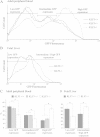
Figure 1
Figure 1
Loss of KLF3 results in up-regulation of the human α-globin gene in a transgenic mouse model. Line3 mice, containing a GFP transgene under the control of the human α-globin proximal promoter and HS-40 enhancer [22], were crossed with Klf3+/− mice to generate Line3::Klf3+/+, Line3::Klf3+/− and Line3::Klf3−/− mice, all homozygous for the transgene. Erythroid GFP fluorescence was then measured by flow cytometry. Shown are representative fluorescence profiles of (A) peripheral blood from mice at 3 weeks of age and (B) TER119+ sorted erythrocytes from embryonic day E14.5 fetal liver. The populations were gated to identify cells expressing low, intermediate and high levels of GFP. Statistical analysis of these gated populations is shown for (C) erythrocytes from mice at 3 weeks of age and (D) TER119+ fetal liver cells. For erythrocytes analyzed at 3 weeks of age, n = 32 for Line3::Klf3+/+, n = 48 for Line3::Klf3+/− and n = 8 for Line3::Klf3−/−. For the analysis of fetal erythrocytes, n = 3 for Line3::Klf3+/+and n = 4 for Line3::Klf3−/−. Error bars represent standard deviation and * represents P < 0.05 (two tailed t-test).
Figure 2
Figure 2
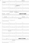
The α-globin promoter contains many consensus KLF3 binding sites. The human HBA2(A) and mouse Hba-a2(B)α-globin proximal promoter sequences, immediately 5′ to the transcriptional start site, were inspected for consensus binding sites, conforming to the sequence 5′-NCN CNC CCN-3′. The position and direction of binding sites are indicated by grey arrows. The sequences used in the design of probes for electrophoretic mobility shift assays are shown by black arrows. Also indicated are CAAT and TATA boxes. Sequences are numbered with respect to the transcription start site at +1.
Figure 3
Figure 3
KLF3 binds the α-globin promoter in vitro. The binding of KLF3 to the α-globin promoter was assessed by EMSA, using radiolabeled probes designed from analysis of the human and mouse α-globin proximal promoter sequences (Figure 2). KLF3 was either expressed in COS-7 cells (lanes 2, 3, 7 and 8) or endogenous KLF3 was harvested in nuclear extracts from K562 (lanes 4 and 5) and MEL (lanes 9 and 10) erythroid cell lines. Nuclear extracts from mock transfected COS-7 cells have been included as a negative control (lanes 1 and 6). Binding to the human promoter sequence is shown in the left hand panel whilst binding to the mouse sequence is shown on the right. αKLF3 indicates an anti-KLF3 antibody used to validate KLF3 specific binding by supershift. Additional bands in lanes 4, 5, 9 and 10 (denoted by asterisks) most likely represent SP1 and SP3 as in [12].
Figure 4
Figure 4
KLF3 binds the human and mouse α-globin promoters in vivo in chromatin immunoprecipitation assays. An anti-KLF3 antibody was used to immunoprecipitate chromatin from the following cell types: (A) uninduced MEL cells, (B) induced MEL cells, (C) mouse primary erythroblasts, (D) uninduced interspecific MEL hybrids containing a normal copy of human chromosome 16, (E) induced interspecific MEL hybrids, and (F) human primary erythroblasts. The y-axis represents enrichment over input DNA, normalized to a control sequence in the Gapdh gene (mouse) or 18S (human). The x-axis represents the positions of the TaqMan probes used. The coding sequence is represented by the three exons (Promoter/Ex1, Ex2, and Ex3) of the α-globin genes. HS- primer sets refer to upstream DNase-hypersensitive regions. Zeta pr refers to the mouse and human embryonic α-globin promoters (Hba-x and HBZ). Inter, refers to the intergenic region (between mouse Hba-a1 and Hba-a2). 5' and 3' are negative controls flanking the α-globin gene. β-actin and β-globin denote control sequences at the β-actin gene and β-globin promoter respectively. Error bars correspond to ±1 standard deviation from at least two independent ChIPs.
Figure 5
Figure 5
α-globin gene expression is de-repressed in murine embryonic fibroblasts lacking KLF3.α-globin mRNA expression levels were determined by real time qRT-PCR analysis of (A) TER119+ erythrocytes purified from embryonic day E14.5 fetal liver (Klf3+/+n = 3, Klf3+/− n = 5, Klf3−/− n = 6), (B) primary MEFs (Klf3+/+n = 2, Klf3−/− n = 2 or 3), (C) immortalized MEFs (Klf3+/+n = 2, Klf3−/− n = 2), and (D) immortalized Klf3−/− MEFs rescued with KLF3-V5 or empty vector (n = 2 for each). (A-D) In each case, relative expression of α-globin mRNA was normalized to 18S rRNA levels, and the expression levels of Klf3+/+(A-C) or Klf3−/−(D) were set to 1.0. In (B), mRNA levels of Klf3 and two known KLF3-repressed targets, Klf8 and Fam132a[7,25], have also been analyzed together with a negative control, Gapdh. In (A-D), error bars shown represent standard error of the mean, *P < 0.05 (one-tailed t-test relative to Klf3+/+), **P < 0.002 (two-tailed t-test relative to Klf3+/+), ***P < 0.05 (two-tailed t-test relative to Klf3−/−). (E) KLF3 ChIP-Seq track across the murine α-globin locus in MEFs from [24]. The positions of the HS-12 and HS-26 regulatory regions are indicated. (F) EMSA showing the binding of KLF3 to two sites within the HS-26 region. Nuclear extracts were obtained from COS-7 cells that were mock-transfected (lanes 1 and 6) or transfected with pMT3-Klf3 (lanes 2, 3, 7 and 8). Nuclear extracts from MEFs are shown in lanes 4, 5, 9 and 10. Identification of KLF3:DNA complexes was achieved by addition of an antibody specific for KLF3 (αKLF3, lanes 3, 5, 8 and 10).
Figure 6
Figure 6
KLF3 from erythroid and non-erythroid cells display similar DNA-binding abilities in vitro. (A) EMSAs were employed to assess the binding of KLF3 to the murine α-globin promoter (lanes 1–7) and a site in the HS-26 element from Figure 5 (lanes 8–14). Nuclear extracts were harvested from non-erythroid MEF (lanes 4, 5, 11 and 12) or erythroid MEL cells (lanes 6, 7, 13 and 14). Nuclear extracts from mock transfected COS-7 cells (lanes 1 and 8) or cells expressing KLF3 (lanes 2, 3, 9 and 10) were included as negative and positive controls respectively. The identity of KLF3 was confirmed by specific antibody supershifts (lanes 3, 5, 7, 10, 12 and 14). (B) Western blot demonstrating the relative amounts of KLF3 in MEF (lane 4) and MEL (lane 5) nuclear extracts used in the EMSAs in (A). As negative and positive controls, COS and COS-KLF3 nuclear extracts have been included (lanes 2 and 3) at 20-fold lower relative amounts than in (A) to facilitate visualization. A size ladder is shown in lane 1.

References

    1. McConnell BB, Yang VW. Mammalian Kruppel-like factors in health and diseases. Physiol Rev. 2010;90(4):1337–1381. doi: 10.1152/physrev.00058.2009. - DOI - PMC - PubMed
    1. Pearson RC, Funnell AP, Crossley M. The mammalian zinc finger transcription factor Kruppel-like factor 3 (KLF3/BKLF) IUBMB life. 2011;63(2):86–93. - PubMed
    1. Pearson R, Fleetwood J, Eaton S, Crossley M, Bao S. Kruppel-like transcription factors: A functional family. Int J Biochem Cell Biol. 2008;40(10):1996–2001. doi: 10.1016/j.biocel.2007.07.018. - DOI - PubMed
    1. Turner J, Crossley M. Cloning and characterization of mCtBP2, a co-repressor that associates with basic Kruppel-like factor and other mammalian transcriptional regulators. Embo J. 1998;17(17):5129–5140. doi: 10.1093/emboj/17.17.5129. - DOI - PMC - PubMed
    1. Funnell AP, Norton LJ, Mak KS, Burdach J, Artuz CM, Twine NA, Wilkins MR, Power CA, Hung TT, Perdomo J, Koh P, Bell-Anderson KS, Orkin SH, Fraser ST, Perkins AC, Pearson RC, Crossley M. The CACCC-binding protein KLF3/BKLF represses a subset of KLF1/EKLF target genes and is required for proper erythroid maturation in vivo. Mol Cell Biol. 2012;32(16):3281–3292. doi: 10.1128/MCB.00173-12. - DOI - PMC - PubMed

Publication types

Substances

LinkOut - more resources