

Evaluation of subcortical grey matter abnormalities in patients with MRI-negative cortical epilepsy determined through structural and tensor magnetic resonance imaging
- PMID: 24885823
- PMCID: PMC4080585
- DOI: 10.1186/1471-2377-14-104
Evaluation of subcortical grey matter abnormalities in patients with MRI-negative cortical epilepsy determined through structural and tensor magnetic resonance imaging
Abstract
Background: Although many studies have found abnormalities in subcortical grey matter (GM) in patients with temporal lobe epilepsy or generalised epilepsies, few studies have examined subcortical GM in focal neocortical seizures. Using structural and tensor magnetic resonance imaging (MRI), we evaluated subcortical GM from patients with extratemporal lobe epilepsy without visible lesion on MRI. Our aims were to determine whether there are structural abnormalities in these patients and to correlate the extent of any observed structural changes with clinical characteristics of disease in these patients.
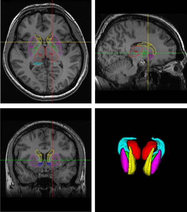
Methods: Twenty-four people with epilepsy and 29 age-matched normal subjects were imaged with high-resolution structural and diffusion tensor MR scans. The patients were characterised clinically by normal brain MRI scans and seizures that originated in the neocortex and evolved to secondarily generalised convulsions. We first used whole brain voxel-based morphometry (VBM) to detect density changes in subcortical GM. Volumetric data, values of mean diffusivity (MD) and fractional anisotropy (FA) for seven subcortical GM structures (hippocampus, caudate nucleus, putamen, globus pallidus, nucleus accumbens, thalamus and amygdala) were obtained using a model-based segmentation and registration tool. Differences in the volumes and diffusion parameters between patients and controls and correlations with the early onset and progression of epilepsy were estimated.
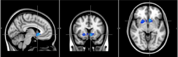
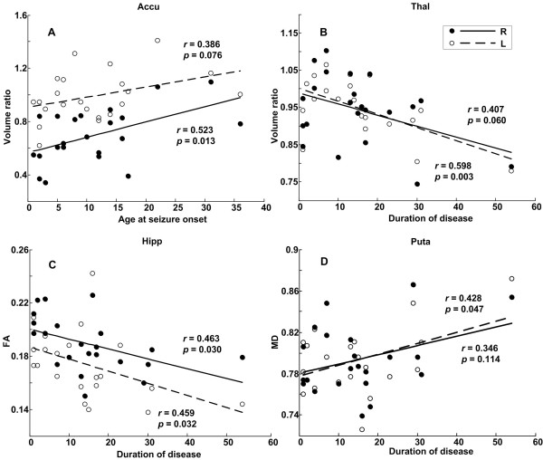
Results: Reduced volumes and altered diffusion parameters of subcortical GM were universally observed in patients in the subcortical regions studied. In the patient-control group comparison of VBM, the right putamen, bilateral nucleus accumbens and right caudate nucleus of epileptic patients exhibited a significantly decreased density Segregated volumetry and diffusion assessment of subcortical GM showed apparent atrophy of the left caudate nucleus, left amygdala and right putamen; reduced FA values for the bilateral nucleus accumbens; and elevated MD values for the left thalamus, right hippocampus and right globus pallidus A decreased volume of the nucleus accumbens consistently related to an early onset of disease. The duration of disease contributed to the shrinkage of the left thalamus.
Conclusions: Patients with neocortical seizures and secondary generalisation had smaller volumes and microstructural anomalies in subcortical GM regions. Subcortical GM atrophy is relevant to the early onset and progression of epilepsy.
Figures
Similar articles
-
Subcortical grey matter changes in juvenile myoclonic epilepsy.Neuroimage Clin. 2017 Nov 3;17:397-404. doi: 10.1016/j.nicl.2017.11.001. eCollection 2018. Neuroimage Clin. 2017. PMID: 29159052 Free PMC article.
-
Cortical-Subcortical morphometric signature of hot water epilepsy patients.Epilepsy Res. 2020 Nov;167:106436. doi: 10.1016/j.eplepsyres.2020.106436. Epub 2020 Aug 8. Epilepsy Res. 2020. PMID: 32846313
-
Detection of structural abnormalities of cortical and subcortical gray matter in patients with MRI-negative refractory epilepsy using neurite orientation dispersion and density imaging.Phys Med. 2018 Apr;48:47-54. doi: 10.1016/j.ejmp.2018.03.005. Epub 2018 Mar 28. Phys Med. 2018. PMID: 29728228
-
Subcortical Alterations in Newly Diagnosed Epilepsy and Associated Changes in Brain Connectivity and Cognition.Hum Brain Mapp. 2024 Nov;45(16):e70069. doi: 10.1002/hbm.70069. Hum Brain Mapp. 2024. PMID: 39508641 Free PMC article. Review.
-
Grey matter volume alterations in trigeminal neuralgia: A systematic review and meta-analysis of voxel-based morphometry studies.Prog Neuropsychopharmacol Biol Psychiatry. 2020 Mar 2;98:109821. doi: 10.1016/j.pnpbp.2019.109821. Epub 2019 Nov 19. Prog Neuropsychopharmacol Biol Psychiatry. 2020. PMID: 31756417
Cited by
-
Subcortical Volume Changes in Migraine with Aura.J Clin Neurol. 2019 Oct;15(4):448-453. doi: 10.3988/jcn.2019.15.4.448. J Clin Neurol. 2019. PMID: 31591831 Free PMC article.
-
Preoperative evaluation of dynamic contrast-enhanced MRI-guided lesion identification using morphometric and textural analysis for patients with epilepsy.Exp Ther Med. 2020 Nov;20(5):98. doi: 10.3892/etm.2020.9228. Epub 2020 Sep 17. Exp Ther Med. 2020. PMID: 32973947 Free PMC article.
-
Multimodal quantitative magnetic resonance imaging analysis with individualized postprocessing in patients with drug-resistant focal epilepsy and conventional visual inspection negative for epileptogenic lesions.Clinics (Sao Paulo). 2019;74:e908. doi: 10.6061/clinics/2019/e908. Epub 2019 Jul 22. Clinics (Sao Paulo). 2019. PMID: 31340255 Free PMC article.
-
Grey matter volume in healthy and epileptic beagles using voxel-based morphometry - a pilot study.BMC Vet Res. 2018 Feb 20;14(1):50. doi: 10.1186/s12917-018-1373-8. BMC Vet Res. 2018. PMID: 29463250 Free PMC article.
-
Limbic-Auditory Interactions of Tinnitus: An Evaluation Using Diffusion Tensor Imaging.Clin Neuroradiol. 2017 Jun;27(2):221-230. doi: 10.1007/s00062-015-0473-0. Epub 2015 Oct 21. Clin Neuroradiol. 2017. PMID: 26490370
References
-
- Saini J, Sinha S, Bagepally BS, Ramchandraiah CT, Thennarasu K, Prasad C, Taly AB, Satishchandra P. Subcortical structural abnormalities in juvenile myoclonic epilepsy (JME): MR volumetry and vertex based analysis. Seizure. 2013;22:230–235. - PubMed
-
- Luo C, Xia Y, Li Q, Xue K, Lai Y, Gong Q, Zhou D, Yao D. Diffusion and volumetry abnormalities in subcortical nuclei of patients with absence seizures. Epilepsia. 2011;52:1092–1099. - PubMed
-
- Du H, Zhang Y, Xie B, Wu N, Wu G, Wang J, Jiang T, Feng H. Regional atrophy of the basal ganglia and thalamus in idiopathic generalized epilepsy. J Magn Reson Imaging. 2011;33:817–821. - PubMed
Publication types
MeSH terms
LinkOut - more resources
Full Text Sources
Other Literature Sources
Medical
