

Efficacy of adalimumab in young children with juvenile idiopathic arthritis and chronic uveitis: a case series
- PMID: 24886032
- PMCID: PMC4045933
- DOI: 10.1186/1756-0500-7-316
Efficacy of adalimumab in young children with juvenile idiopathic arthritis and chronic uveitis: a case series
Abstract
Background: Juvenile idiopathic arthritis is a relatively common chronic disease of childhood, and is associated with persistent morbidity and extra-articular complications, one of the most common being uveitis. The introduction of biologic therapies, particularly those blocking the inflammatory mediator tumor necrosis factor-α, provided a new treatment option for juvenile idiopathic arthritis patients who were refractory to standard therapy such as non-steroidal anti-inflammatory drugs, corticosteroids and/or methotrexate.
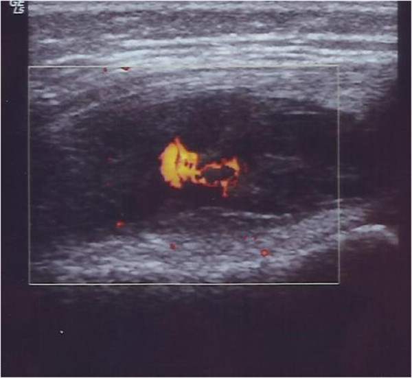
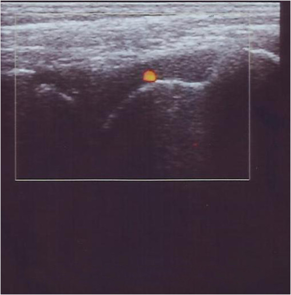
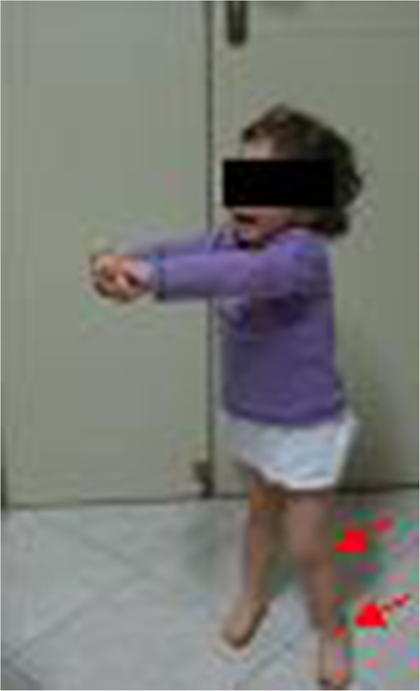
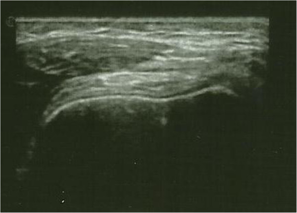
Case presentations: The first case was a 2-year-old girl with juvenile idiopathic arthritis and uveitis who failed to respond to treatment with anti-inflammatories, low-dose corticosteroids and methotrexate, and had growth retardation. Adalimumab 24 mg/m2 every 2 weeks and prednisone 0.5 mg/kg/day were added to methotrexate therapy; steroid tapering and withdrawal started after 1 month. After 2 months the patient showed good control of articular and ocular manifestations, and she remained in remission for 1 year, receiving adalimumab and methotrexate with no side effects, and showing significant improvement in growth. Case 2 was a 9-year-old boy with an 8-year history of juvenile idiopathic arthritis and uveitis that initially responded to infliximab, but relapse occurred after 2 years off therapy. After switching to adalimumab, and adjusting doses of both adalimumab and methotrexate based on body surface area, the patient showed good response and corticosteroids were tapered and withdrawn after 6 months; the patient remained in remission taking adalimumab and methotrexate. The final case was a 5-year-old girl with juvenile idiopathic arthritis for whom adalimumab was added to methotrexate therapy after three flares of uveitis. The patient had two subsequent episodes of uveitis that responded well to local therapy, but was then free of both juvenile idiopathic arthritis and uveitis symptoms, allowing methotrexate and then adalimumab to be stopped; the patient remained in drug-free remission.
Conclusion: This report includes the first published case of the use of adalimumab in a child aged <3 years. Our clinical experience further supports the use of biologic therapy for the management of juvenile idiopathic arthritis and uveitis in children as young as two years of age.
Figures
Similar articles
-
Adalimumab in combination with methotrexate for refractory uveitis associated with juvenile idiopathic arthritis: a RCT.Health Technol Assess. 2019 Apr;23(15):1-140. doi: 10.3310/hta23150. Health Technol Assess. 2019. PMID: 31033434 Free PMC article. Clinical Trial.
-
A randomised controlled trial of the clinical effectiveness, safety and cost-effectiveness of adalimumab in combination with methotrexate for the treatment of juvenile idiopathic arthritis associated uveitis (SYCAMORE Trial).Trials. 2014 Jan 9;15:14. doi: 10.1186/1745-6215-15-14. Trials. 2014. PMID: 24405833 Free PMC article. Clinical Trial.
-
Adalimumab in the therapy of uveitis in childhood.Br J Ophthalmol. 2007 Mar;91(3):319-24. doi: 10.1136/bjo.2006.103721. Epub 2006 Oct 11. Br J Ophthalmol. 2007. PMID: 17035274 Free PMC article.
-
Juvenile idiopathic arthritis-associated uveitis.Best Pract Res Clin Rheumatol. 2017 Aug;31(4):517-534. doi: 10.1016/j.berh.2018.01.002. Epub 2018 Feb 26. Best Pract Res Clin Rheumatol. 2017. PMID: 29773271 Review.
-
Anti-TNF therapy for juvenile idiopathic arthritis-related uveitis.Drug Des Devel Ther. 2014 Mar 24;8:341-8. doi: 10.2147/DDDT.S54207. eCollection 2014. Drug Des Devel Ther. 2014. PMID: 24711694 Free PMC article. Review.
Cited by
-
Biologic Therapies in Sarcoidosis and Uveitis: A Review.Cureus. 2020 Jul 7;12(7):e9057. doi: 10.7759/cureus.9057. Cureus. 2020. PMID: 32782876 Free PMC article. Review.
-
Outcome of Juvenile Idiopathic Arthritis Associated Uveitis in Two Disease Subtypes.Arch Rheumatol. 2017 Jan 6;32(1):26-31. doi: 10.5606/ArchRheumatol.2017.6060. eCollection 2017 Mar. Arch Rheumatol. 2017. PMID: 30375520 Free PMC article.
References
-
- Petty RE, Southwood TR, Manners P, Baum J, Glass DN, Goldenberg J, He X, Maldonado-Cocco J, Orozco-Alcala J, Prieur AM, Suarez-Almazor ME, Woo P. International League of Associations for Rheumatology. International League of Associations for Rheumatology classification of juvenile idiopathic arthritis: second revision, Edmonton, 2001. J Rheumatol. 2004;31:390–392. - PubMed
-
- Petty RE, Southwood TR, Baum J, Bhettay E, Glass DN, Manners P, Maldonado-Cocco J, Suarez-Almazor M, Orozco-Alcala J, Prieur AM. Revision of the proposed classification criteria for juvenile idiopathic arthritis: Durban, 1997. J Rheumatol. 1998;25(10):1991–1994. - PubMed
-
- Gaspari S, Marcovecchio ML, Breda L, Chiarelli F. Growth in juvenile idiopathic arthritis: the role of inflammation. Clin Exp Rheumatol. 2011;29(1):104–110. - PubMed
Publication types
MeSH terms
Substances
LinkOut - more resources
Full Text Sources
Other Literature Sources
Medical
