. 2014 May 15:11:89.
doi: 10.1186/1743-422X-11-89.

Viral metagenomic analysis of feces of wild small carnivores

Affiliations

Viral metagenomic analysis of feces of wild small carnivores

Rogier Bodewes et al. Virol J. .

Abstract

Background: Recent studies have clearly demonstrated the enormous virus diversity that exists among wild animals. This exemplifies the required expansion of our knowledge of the virus diversity present in wildlife, as well as the potential transmission of these viruses to domestic animals or humans.

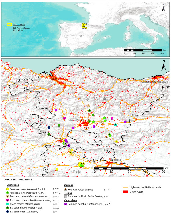
Methods: In the present study we evaluated the viral diversity of fecal samples (n = 42) collected from 10 different species of wild small carnivores inhabiting the northern part of Spain using random PCR in combination with next-generation sequencing. Samples were collected from American mink (Neovison vison), European mink (Mustela lutreola), European polecat (Mustela putorius), European pine marten (Martes martes), stone marten (Martes foina), Eurasian otter (Lutra lutra) and Eurasian badger (Meles meles) of the family of Mustelidae; common genet (Genetta genetta) of the family of Viverridae; red fox (Vulpes vulpes) of the family of Canidae and European wild cat (Felis silvestris) of the family of Felidae.

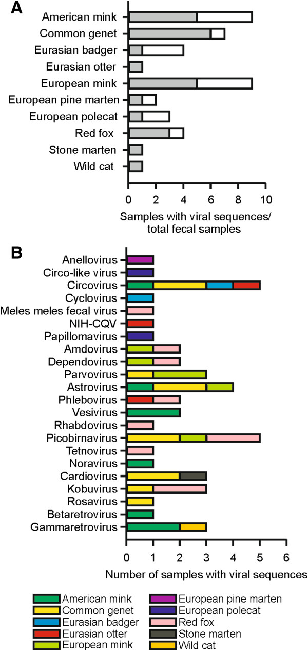
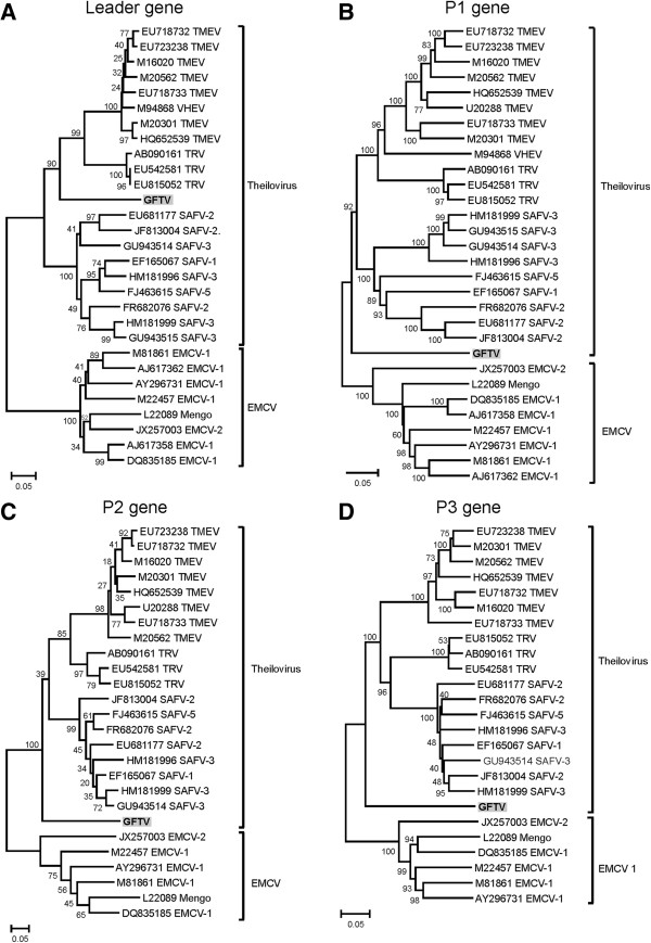
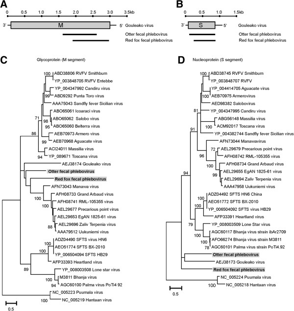
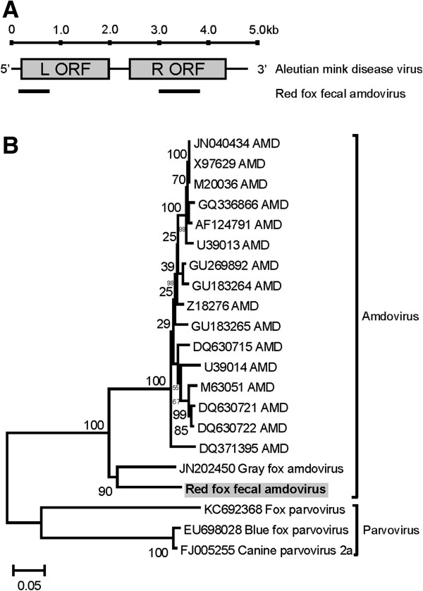
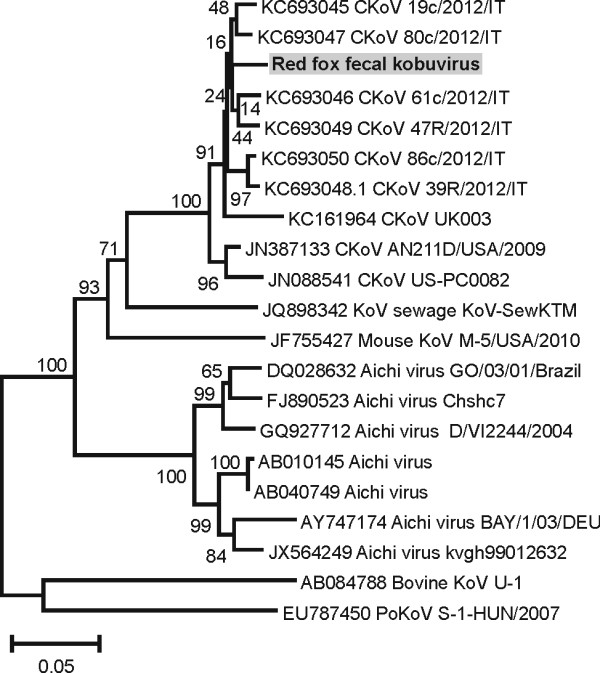
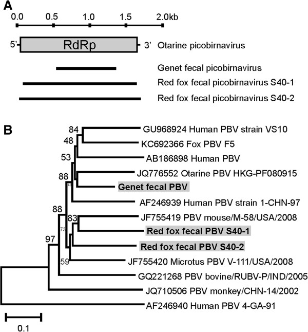
Results: A number of sequences of possible novel viruses or virus variants were detected, including a theilovirus, phleboviruses, an amdovirus, a kobuvirus and picobirnaviruses.

Conclusions: Using random PCR in combination with next generation sequencing, sequences of various novel viruses or virus variants were detected in fecal samples collected from Spanish carnivores. Detected novel viruses highlight the viral diversity that is present in fecal material of wild carnivores.


Figures

Figure 1
Figure 1
Map with locations of sample collection. Maps showing the location of the study area in the Basque Country and La Rioja regions (Spain) and spatial distribution of the 42 analysed carnivore samples of the Mustelidae, Canidae, Felidae and Viverridae families.
Figure 2
Figure 2
Overview of detected viral sequences in the present study. Number of animals included in this study (bars) and the number of samples in which viruses where detected (grey areas; A). Number of animals in which viral sequences were detected for each genus/ group (B).
Figure 3
Figure 3
Phylogenetic analysis of Genet fecal theilovirus (GFTV). Phylogenetic neighbor-joining tree with p-distance and 1,000 bootstrap replicates of the nucleotide sequence of the Leader gene (A), P1 gene (B), P2 gene (C) and partial P3 gene (D) of Genet fecal theiloviruses and various other viruses of the genus Cardiovirus. TMEV: Theiler’s murine ecephalomyelitis vrus, VHEV: Vilyuisk human encephalomyelitis virus, TRV: Theiler’s-like rat virus, SAFV: Saffold virus, EMCV: encephalomyocarditis virus. Indicated are genbank accession numbers and bootstrap values.
Figure 4
Figure 4
Phylogenetic analysis of Red fox fecal phlebovirus and Otter fecal phlebovirus. Overview of obtained viral sequence of the M segment (A) and S segment (B) of Otter fecal phlebovirus and Red fox fecal phlebovirus using Gouleako virus as a reference. Phylogenetic maximum likelihood tree (WAG + F + I + G model) with 100 bootstrap replicates of the deduced amino acid sequence of the partial glycoprotein gene (C) and nucleoprotein gene (D) of Red fox and Otter fecal phleboviruses and various other viruses of the genus Phlebovirus. RVFV: Rift valley fever virus, SFTS: Severe fever with thrombocytopenia syndrome virus. Indicated are Genbank accession numbers. Only bootstrap values above 70 are indicated.
Figure 5
Figure 5
Phylogenetic analysis of Red fox fecal amdovirus. Overview of obtained viral sequences of Red fox fecal amdovirus using Aleutian mink disease virus as a reference (A). Phylogenetic neighbor-joining tree with p-distance and 1,000 bootstrap replicates of the deduced amino acid sequence of the partial VP2 gene of Red fox fecal amdovirus and VP2 genes of various other viruses of the genus Amdovirus detected in mink and gray foxes and viruses of the genus Parvovirus detected in red and blue foxes and dogs (B). Indicated are Genbank accession numbers and bootstrap values.
Figure 6
Figure 6
Phylogenetic analysis of Red fox fecal kobuvirus S37. Phylogenetic neighbor-joining tree with p-distance and 1,000 bootstrap replicates of the nucleotide sequence of the partial 3D gene of Red fox fecal kobuvirus and partial 3D genes of various other viruses of the genus Kobuvirus. Indicated are Genbank accession numbers and bootstrap values. CKoV: Canine kobuvirus, KoV: kobuvirus, PoKoV: Porcine kobuvirus.
Figure 7
Figure 7
Phylogenetic analysis of Genet fecal picobirnavirus and Red fox fecal picobirnavirus S40-1 and S40-2. Overview of obtained viral sequences of Genet fecal picobirnavirus and Red fox fecal picobirnavirus S40-1 and S40-2 using segment 2 of Otarine picobirnavirus as a reference (A). Phylogenetic neighbor-joining tree with p-distance and 1,000 bootstrap replicates of the deduced amino acid sequence of the RdRp gene of Red fox fecal picobirnavirus S40-1, S40-2 and Genet fecal picobirnavirus and near complete RdRp genes of various other picobirnaviruses. Indicated are Genbank accession numbers and bootstrap values. PBV: picobirnavirus (B).

Similar articles

Cited by

References

    1. Zaki AM, van Boheemen S, Bestebroer TM, Osterhaus AD, Fouchier RA. Isolation of a novel coronavirus from a man with pneumonia in Saudi Arabia. N Engl J Med. 2012;367:1814–1820. doi: 10.1056/NEJMoa1211721. - DOI - PubMed
    1. Reusken CB, Haagmans BL, Müller MA, Gutierrez C, Godeke GJ, Meyer B, Muth D, Raj VS, Smits-De Vries L, Corman VM, Drexler JF, Smits SL, El Tahir YE, De Sousa R, van Beek J, Nowotny N, van Maanen K, Hidalgo-Hermoso E, Bosch BJ, Rottier P, Osterhaus A, Gortázar-Schmidt C, Drosten C, Koopmans MP. Middle East respiratory syndrome coronavirus neutralising serum antibodies in dromedary camels: a comparative serological study. Lancet Infect Dis. 2013;13:859–866. doi: 10.1016/S1473-3099(13)70164-6. - DOI - PMC - PubMed
    1. Ithete NL, Stoffberg S, Corman VM, Cottontail VM, Richards LR, Schoeman MC, Drosten C, Drexler JF, Preiser W. Close relative of human Middle East respiratory syndrome coronavirus in bat, South Africa. Emerg Infect Dis. 2013;19:1697–1699. doi: 10.3201/eid1910.130946. - DOI - PMC - PubMed
    1. Gao R, Cao B, Hu Y, Feng Z, Wang D, Hu W, Chen J, Jie Z, Qiu H, Xu K, Xu X, Lu H, Zhu W, Gao Z, Xiang N, Shen Y, He Z, Gu Y, Zhang Z, Yang Y, Zhao X, Zhou L, Li X, Zou S, Zhang Y, Li X, Yang L, Guo J, Dong J, Li Q. et al.Human infection with a novel avian-origin influenza A (H7N9) virus. N Engl J Med. 2013;368:1888–1897. doi: 10.1056/NEJMoa1304459. - DOI - PubMed
    1. Liu D, Shi W, Shi Y, Wang D, Xiao H, Li W, Bi Y, Wu Y, Li X, Yan J, Liu W, Zhao G, Yang W, Wang Y, Ma J, Shu Y, Lei F, Gao GF. Origin and diversity of novel avian influenza A H7N9 viruses causing human infection: phylogenetic, structural, and coalescent analyses. Lancet. 2013;381:1926–1932. doi: 10.1016/S0140-6736(13)60938-1. - DOI - PubMed

Publication types