. 2014 May 13:11:35.
doi: 10.1186/1742-4690-11-35.

Rapid inflammasome activation in microglia contributes to brain disease in HIV/AIDS

Affiliations

Rapid inflammasome activation in microglia contributes to brain disease in HIV/AIDS

John G Walsh et al. Retrovirology. .

Abstract

Background: Human immunodeficiency virus type 1(HIV-1) infects and activates innate immune cells in the brain resulting in inflammation and neuronal death with accompanying neurological deficits. Induction of inflammasomes causes cleavage and release of IL-1β and IL-18, representing pathogenic processes that underlie inflammatory diseases although their contribution HIV-associated brain disease is unknown.

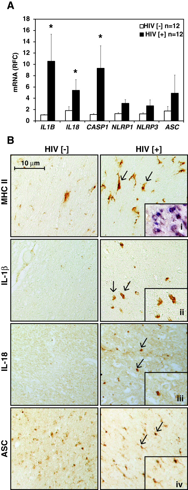
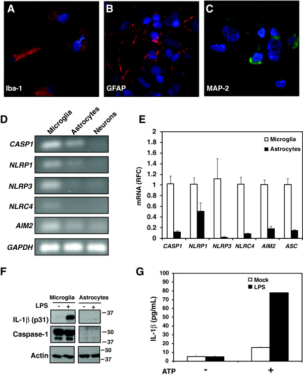
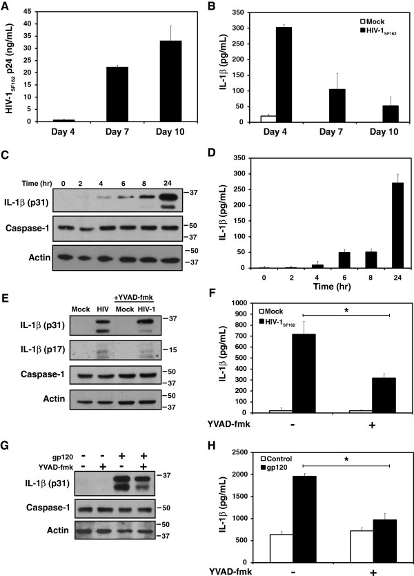
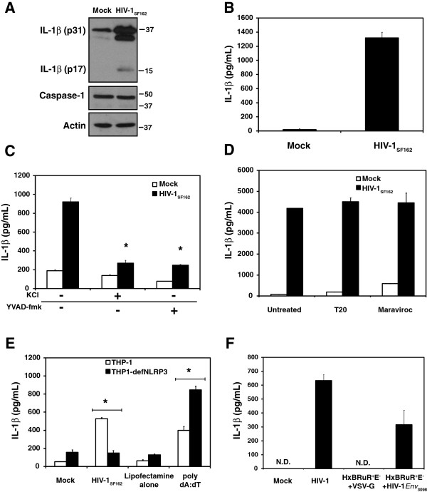
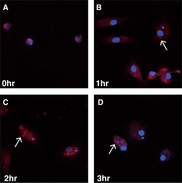
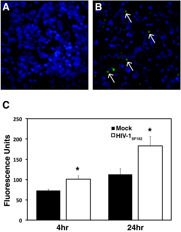
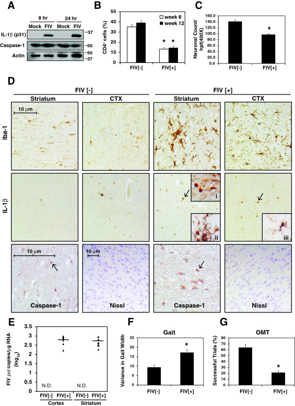
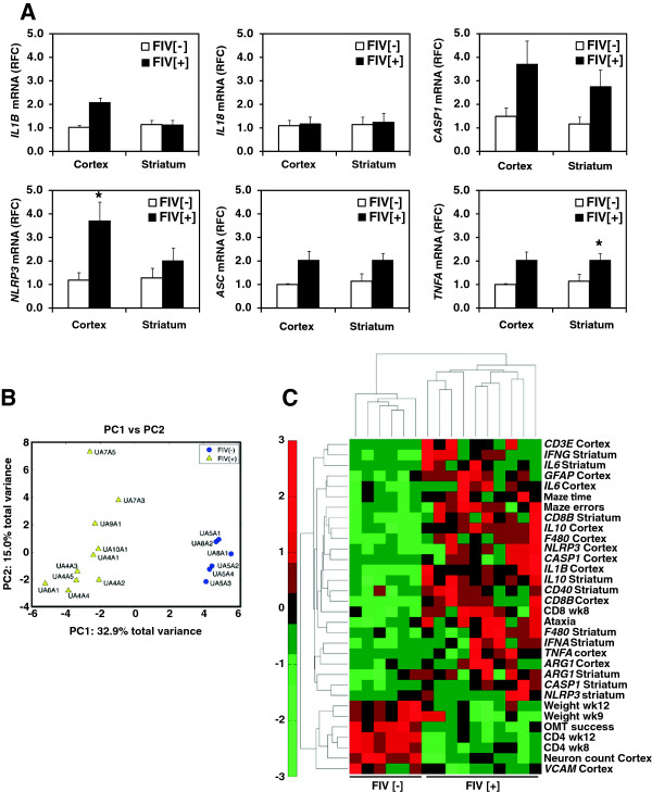
Results: Investigation of inflammasome-associated genes revealed that IL-1β, IL-18 and caspase-1 were induced in brains of HIV-infected persons and detected in brain microglial cells. HIV-1 infection induced pro-IL-1β in human microglia at 4 hr post-infection with peak IL-1β release at 24 hr, which was accompanied by intracellular ASC translocation and caspase-1 activation. HIV-dependent release of IL-1β from a human macrophage cell line, THP-1, was inhibited by NLRP3 deficiency and high extracellular [K+]. Exposure of microglia to HIV-1 gp120 caused IL-1β production and similarly, HIV-1 envelope pseudotyped viral particles induced IL-1β release, unlike VSV-G pseudotyped particles. Infection of cultured feline macrophages by the related lentivirus, feline immunodeficiency virus (FIV), also resulted in the prompt induction of IL-1β. In vivo FIV infection activated multiple inflammasome-associated genes in microglia, which was accompanied by neuronal loss in cerebral cortex and neurological deficits. Multivariate analyses of data from FIV-infected and uninfected animals disclosed that IL-1β, NLRP3 and caspase-1 expression in cerebral cortex represented key molecular determinants of neurological deficits.

Conclusions: NLRP3 inflammasome activation was an early and integral aspect of lentivirus infection of microglia, which was associated with lentivirus-induced brain disease. Inflammasome activation in the brain might represent a potential target for therapeutic interventions in HIV/AIDS.


Figures

Figure 1
Figure 1
Expression of inflammasome components and substrates in the HIV-1 infected human brain. A. Relative fold change (RFC) in mRNA expression in the white matter of persons with HIV-1 infection (HIV [+]) (n = 12) compared to other disease controls (HIV [-]) (n = 12), as measured by semi-quantitative real-time PCR. Mean values are reported. Bars indicate standard error. *indicate p-value of <0.05. B. Immunohistochemistry staining of cerebral white matter sections from other disease control (HIV [-]) and HIV-infected (HIV [+]) (200X view). Inset 'i’ represents double immunostaining for MHC II and IL-1β. Insets 'ii’, 'iii’ and 'iv’ are magnified images of the indicated cells within the HIV [+] section.
Figure 2
Figure 2
Expression and activation of the inflammasome in primary human CNS cells. Isolated primary human cells including microglia (A), astrocytes (B) and neurons (C) were immunolabelled with antibodies to Iba-1, GFAP and MAP-2, respectively, to verify the purity of cell cultures. D. Qualitative RT-PCR showing transcript expression of caspase-1 as well as various inflammasome-forming cytoplasmic pattern recognition receptors in primary cultured human microglia, astrocytes and neurons. E. Semi-quantitative real-time PCR showing the relative fold change in inflammasome-associated gene expression in cultured primary human microglia and astrocytes. Bars indicate standard error. F. Protein expression of IL-1β and caspase-1 in primary human microglia but not astrocytes. G. IL-1β release from Mock-or LPS-primed (10 ng/mL, 6 hr) primary human microglia followed by ATP stimulation (5 mM, 1 hr). Mean values are reported. Bars indicate standard error.
Figure 3
Figure 3
IL-1β expression and release from primary human microglia following HIV-1 infection or exposure. A. Viral p24 and B. IL-1β release from microglia at days 4, 7 and 10 following infection with HIV-1SF162.C. IL-1β induction and D. release from microglia cultures over 24 hr following exposure to HIV-1SF162. E. Induction, processing and F. release of IL-1β following 24 hr exposure to HIV-1SF162 +/- the caspase inhibitor YVAD-fmk (80 μM). G. Induction and H. release of IL-1β from microglia cultures exposed to recombinant gp120 (80 nM) for 24 hr +/- YVAD-fmk. Mean levels are reported for each ELISA. Bars indicate standard error. We compared the effect of HIV-1gp120 exposure on IL-1β release +/- YVAD-fmk using two-tailed Student t-test (*p < 0.05).
Figure 4
Figure 4
IL-1β release in response to HIV-1SF162 exposure is dependent on the NLRP3 inflammasome but not viral fusion or CCR5. A. Induction, processing and B. release of IL-1β from PMA-differentiated THP-1 cells following 4 hr exposure to HIV-1SF162. C. IL-1β release from PMA-differentiated THP-1 cells exposed to HIV-1SF162 for 4 hr +/- extracellular KCl (50 mM) or YVAD-fmk (80 μM). D. Release of IL-1β from PMA-differentiated THP-1 cells following 18 hr exposure to HIV-1SF162 +/- the inhibitor of viral fusion T20 (20 μg/mL) or the CCR5 antagonist maraviroc (100nM). E. IL-1β release from PMA-differentiated THP-1 cells or THP1-defNLRP3 cells exposed to HIV-1SF162 or poly dA:dT for 4 hr (poly dA:dT was transfected using Lipofectamine 2000). F. IL-1β release from PMA-differentiated THP-1 cells exposed to HIV-1YU2, HxBRuR+E- + VSVG, or HxBRuR+E- + HIV-1Env3098 for 24 hours. Mean levels are reported for each ELISA. Bars indicate standard error. (*p < 0.05). “N.D.” indicates that IL-1β was “Not Detected”.
Figure 5
Figure 5
HIV-1SF162 infection of human microglia induces rapid ASC intracellular translocation. A. Mock-infected microglia exhibit minimal ASC immunoreactivity. B. At 1 hr post-infection ASC immunoreactivity (red) was detected in the cytoplasm. C. At 2 hr post-infection, punctate ASC immunoreactivity was evident in the cytoplasm of microglia. D. At 3 hr post-infection, aggregates of ASC immunoreactivity was readily apparent in microglia. (Original magnification 400X).
Figure 6
Figure 6
HIV-1SF162 infection of human microglia activates caspase-1. A. Caspase-1 activity was not detected in mock-infected microglial cultures that were labeled with Hoescht nuclear stain. B. In contrast, microglia infected by HIV-1 showed caspase-1 activity at 24 hr post-infection (Original magnification 400X). C. Quantitation of caspase-1 activity in microglia at 4 and 24 hr post-infection showed an increased in caspase-1 activity induced by HIV-1 infection and also over time. (Mean values reported. Bars indicate standard error. Student t-test, *p < 0.05).
Figure 7
Figure 7
IL-1β and caspase-1 expression in the brains of FIV-infected animals with neurologic defects. A. IL-1β immunoreactivity was evident in feline monocyte-derived macrophages at 8 and 24 hr following FIV infection while caspase-1 expression was unaffected. B. CD4+ T cell count in blood of FIV-infected (n = 10) and uninfected animals (n = 6) at weeks 8 and 12 post-infection. C. Neuronal counts per high power field (400X) from FIV [+] animals versus FIV [-] controls at week 12 D. Immunohistochemical detection of Iba-1, IL-1β and caspase-1 in both striatum and cortex (CTX) from FIV [-] and FIV [+] animals (original magnification: Iba-1 and IL-1β X200; caspase-1 X600; Nissl stain, X100). Inset 'i’ shows an IL-1β immunoreactive microglial nodule. Insets 'ii’ and 'iii’ display IL-1β and Iba-1 co-immunolabeling. E. Detection of FIV pol RNA in the cortex and striatum of FIV [-] (n = 6) and FIV [+] animals (n = 10) at week 12. Performance of FIV [-] (n = 6) and FIV [+] (n = 10) animals in neurobehavioral tests of gait (F) and memory (G) at week 12 post-infection. “N.D.” indicates that virus was “Not Detected”.
Figure 8
Figure 8
In vivo association of inflammasome-related genes with neurological disease in FIV [+] animals. A. Relative fold change (RFC) in mRNA expression of inflammasome- related and immune genes in the cortex and striatum of FIV [+] (n = 6) and FIV [-] (n = 10) animals at 12 weeks post-infection. Mean RFC values are reported. Bars indicate standard error. *indicate p-value of <0.05. B. Principal component analysis (PCA) comparing 49 clinical, neurobehavioral and molecular variables in FIV [-] and FIV [+] animals. PC1 represents the separation of the FIV [-] and FIV [+] animals while PC2 represents intra-group variance. C. Hierarchical Cluster Analysis of multivariate similarities between variables and animals. Only variables that significantly contributed to PC1 (infection status) are presented in the heat map.

Similar articles

Cited by

References

    1. Burdo TH, Lackner A, Williams KC. Monocyte/macrophages and their role in HIV neuropathogenesis. Immunol Rev. 2013;254(1):102–113. doi: 10.1111/imr.12068. - DOI - PMC - PubMed
    1. Zink MC, Laast VA, Helke KL, Brice AK, Barber SA, Clements JE, Mankowski JL. From mice to macaques–animal models of HIV nervous system disease. Curr HIV Res. 2006;4(3):293–305. doi: 10.2174/157016206777709410. - DOI - PubMed
    1. Ellis R, Langford D, Masliah E. HIV and antiretroviral therapy in the brain: neuronal injury and repair. Nat Rev Neurosci. 2007;8(1):33–44. doi: 10.1038/nrn2040. - DOI - PubMed
    1. Vivithanaporn P, Heo G, Gamble J, Krentz HB, Hoke A, Gill MJ, Power C. Neurologic disease burden in treated HIV/AIDS predicts survival: a population-based study. Neurology. 2010;75(13):1150–1158. doi: 10.1212/WNL.0b013e3181f4d5bb. - DOI - PMC - PubMed
    1. Mothobi NZ, Brew BJ. Neurocognitive dysfunction in the highly active antiretroviral therapy era. Curr Opin Infect Dis. 2012;25(1):4–9. doi: 10.1097/QCO.0b013e32834ef586. - DOI - PubMed

MeSH terms