Identification of mesenchymal stem cells and osteogenic factors in bone marrow aspirate and peripheral blood for spinal fusion by flow cytometry and proteomic analysis
- PMID: 24886437
- PMCID: PMC4037761
- DOI: 10.1186/1749-799X-9-32
Identification of mesenchymal stem cells and osteogenic factors in bone marrow aspirate and peripheral blood for spinal fusion by flow cytometry and proteomic analysis
Abstract
Background: An in vivo animal study and a prospective clinical study have indicated that bone marrow aspirate (BMA) augments spinal arthrodesis. However, there is no quantified data to explain why fusion rate can be augmented by BMA in lumbar posterolateral fusion.
Methods: To analyze the proportion of mesenchymal stem cells (MSCs) and osteogenic factors in human BMA and peripheral blood (PB) of the same patient. Autologous BMA and PB from the patients were analyzed by flow cytometry (FACS) using cell markers for MSCs. The osteogenic potential of MSCs was determined by alkaline phosphatase (ALP) activity and calcium level quantification. Proteomics were used for the qualitative and quantitative mapping of the whole proteome from BMA and PB plasma. The mass-to-charge ratio was calculated by time-of-flight mass spectrometry (TOF-MS). The overexpression of protein was confirmed using Western blot analysis.
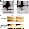
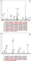
Results: The proportion of MSCs (CD34-/CD29+/CD105+) was higher in the BMA than that in the PB. Colony-forming cell (CFC) assays suggested that fewer colonies were formed in PB cultures than in BMA culture. There was no significant difference in the osteogenic potential of the MSCs between the PB and BMA. Proteomic mass spectrometry assays suggested that the levels of catalase (osteoclast inhibitor) and glutathione peroxidase 3 (osteogenic biomarker) were higher in the BMA than those in the PB, and this was confirmed by Western blot analysis.
Conclusions: The proportions of MSCs and osteogenic factors were higher in the BMA than in the PB. This may explain why fusion rate can be augmented by BMA in lumbar posterolateral fusion.
Figures


Similar articles
-
A comparison of posterolateral lumbar fusion comparing autograft, autogenous laminectomy bone with bone marrow aspirate, and calcium sulphate with bone marrow aspirate: a prospective randomized study.Spine (Phila Pa 1976). 2009 Dec 1;34(25):2715-9. doi: 10.1097/BRS.0b013e3181b47232. Spine (Phila Pa 1976). 2009. PMID: 19940728 Clinical Trial.
-
Clinical and radiographic outcomes of concentrated bone marrow aspirate with allograft and demineralized bone matrix for posterolateral and interbody lumbar fusion in elderly patients.Eur Spine J. 2015 Nov;24(11):2567-72. doi: 10.1007/s00586-015-4117-5. Epub 2015 Jul 14. Eur Spine J. 2015. PMID: 26169879
-
Human mesenchymal stem cell proliferation and osteogenic differentiation during long-term ex vivo cultivation is not age dependent.J Bone Miner Metab. 2011 Mar;29(2):224-35. doi: 10.1007/s00774-010-0215-y. Epub 2010 Sep 2. J Bone Miner Metab. 2011. PMID: 20811759
-
Electrospun, synthetic bone void filler promotes human MSC function and BMP-2 mediated spinal fusion.J Biomater Appl. 2020 Oct-Nov;35(4-5):532-543. doi: 10.1177/0885328220937999. Epub 2020 Jul 5. J Biomater Appl. 2020. PMID: 32627633 Free PMC article.
-
Evaluation of posterolateral spinal fusion using mesenchymal stem cells: differences with or without osteogenic differentiation.Spine (Phila Pa 1976). 2007 Oct 15;32(22):2432-6. doi: 10.1097/BRS.0b013e3181573924. Spine (Phila Pa 1976). 2007. PMID: 18090081
Cited by
-
GEORG-SCHMORL-PRIZE OF THE GERMAN SPINE SOCIETY (DWG) 2016: Comparison of in vitro osteogenic potential of iliac crest and degenerative facet joint bone autografts for intervertebral fusion in lumbar spinal stenosis.Eur Spine J. 2017 May;26(5):1408-1415. doi: 10.1007/s00586-017-5020-z. Epub 2017 Mar 21. Eur Spine J. 2017. PMID: 28324211
-
Stromal Fibroblasts Counteract the Caveolin-1-Dependent Radiation Response of LNCaP Prostate Carcinoma Cells.Front Oncol. 2022 Jan 26;12:802482. doi: 10.3389/fonc.2022.802482. eCollection 2022. Front Oncol. 2022. PMID: 35155239 Free PMC article.
-
Caveolin-1 regulates the ASMase/ceramide-mediated radiation response of endothelial cells in the context of tumor-stroma interactions.Cell Death Dis. 2020 Apr 9;11(4):228. doi: 10.1038/s41419-020-2418-z. Cell Death Dis. 2020. PMID: 32273493 Free PMC article.
-
Impact of the Process Variables on the Yield of Mesenchymal Stromal Cells from Bone Marrow Aspirate Concentrate.Bioengineering (Basel). 2022 Jan 29;9(2):57. doi: 10.3390/bioengineering9020057. Bioengineering (Basel). 2022. PMID: 35200410 Free PMC article. Review.
-
Engrafted peripheral blood-derived mesenchymal stem cells promote locomotive recovery in adult rats after spinal cord injury.Am J Transl Res. 2017 Sep 15;9(9):3950-3966. eCollection 2017. Am J Transl Res. 2017. PMID: 28979672 Free PMC article.
References
-
- Nather A. Use of allograft in spinal surgery. Ann Transpl. 1999;4:19–22. - PubMed
Publication types
MeSH terms
LinkOut - more resources
Full Text Sources
Other Literature Sources

