. 2014 May 15:13:108.
doi: 10.1186/1476-4598-13-108.

DUSP3/VHR is a pro-angiogenic atypical dual-specificity phosphatase

Affiliations

DUSP3/VHR is a pro-angiogenic atypical dual-specificity phosphatase

Mathieu Amand et al. Mol Cancer. .

Abstract

Background: DUSP3 phosphatase, also known as Vaccinia-H1 Related (VHR) phosphatase, encoded by DUSP3/Dusp3 gene, is a relatively small member of the dual-specificity protein phosphatases. In vitro studies showed that DUSP3 is a negative regulator of ERK and JNK pathways in several cell lines. On the other hand, DUSP3 is implicated in human cancer. It has been alternatively described as having tumor suppressive and oncogenic properties. Thus, the available data suggest that DUSP3 plays complex and contradictory roles in tumorigenesis that could be cell type-dependent. Since most of these studies were performed using recombinant proteins or in cell-transfection based assays, the physiological function of DUSP3 has remained elusive.

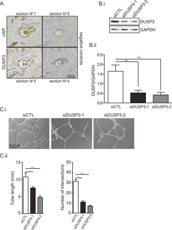
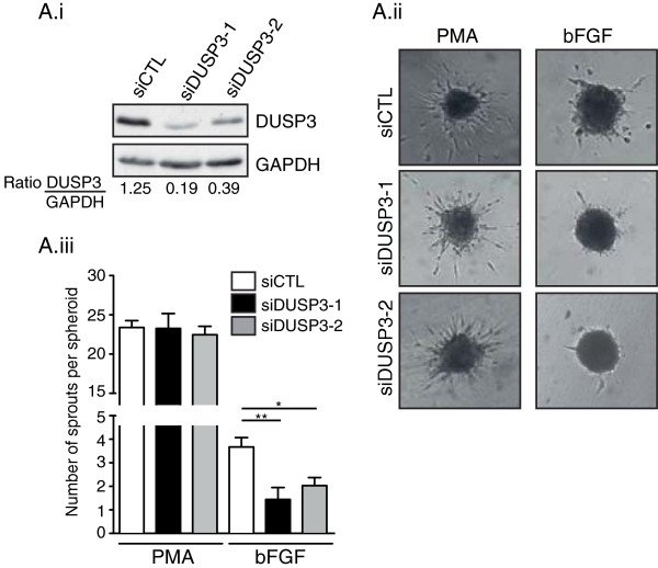
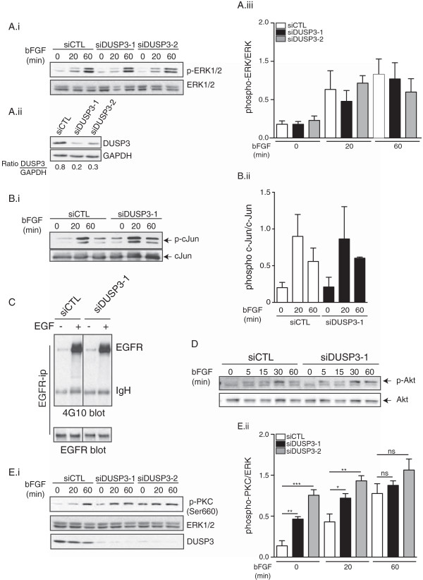
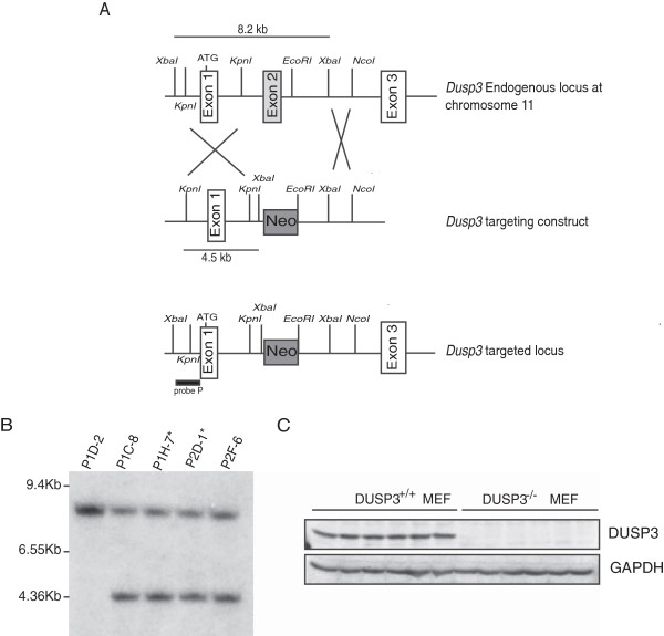
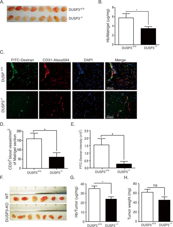
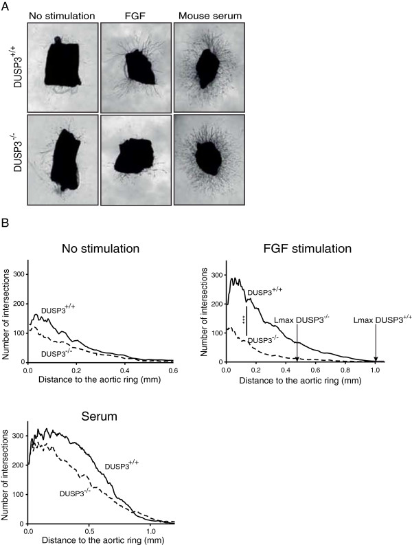
Results: Using immunohistochemistry on human cervical sections, we observed a strong expression of DUSP3 in endothelial cells (EC) suggesting a contribution for this phosphatase to EC functions. DUSP3 downregulation, using RNA interference, in human EC reduced significantly in vitro tube formation on Matrigel and spheroid angiogenic sprouting. However, this defect was not associated with an altered phosphorylation of the documented in vitro DUSP3 substrates, ERK1/2, JNK1/2 and EGFR but was associated with an increased PKC phosphorylation. To investigate the physiological function of DUSP3, we generated Dusp3-deficient mice by homologous recombination. The obtained DUSP3-/- mice were healthy, fertile, with no spontaneous phenotype and no vascular defect. However, DUSP3 deficiency prevented neo-vascularization of transplanted b-FGF containing Matrigel and LLC xenograft tumors as evidenced by hemoglobin (Hb) and FITC-dextran quantifications. Furthermore, we found that DUSP3 is required for b-FGF-induced microvessel outgrowth in the aortic ring assay.

Conclusions: All together, our data identify DUSP3 as a new important player in angiogenesis.


Figures

Figure 1
Figure 1
DUSP3 is highly expressed in endothelial cells and its dowregulation inhibits in vitro tubulogenesis. (A) Immunohistochemistry of DUSP3 and Von Willbrand Factor (vWF) on paraffin embedded 4 μm serial sections of human cervix biopsies. Section 1 was stained with anti-vWF antibody and section 2 with anti-DUSP3 antibody. Sections 3 and 4 were stained with the secondary antibodies used to reveal vWF and DUSP3 staining respectively. (B) HUVEC cells were transfected with non-targeting siRNA (siCTL) or with DUSP3 targeting siRNA (siDUSP3-1 and siDUSP3-2). Efficiency of DUSP3 downregulation was measured at protein level using western blot 72 h after cell transfection. (Bi) Equal amount of proteins were resolved by SDS-PAGE, and western blot was performed using anti-DUSP3 antibody or anti-GAPDH as loading control. (Bii) Quantification and statistical analysis of DUSP3 protein expression in siCTL, siDUSP3-1 and siDUSP3-2 transfection conditions represented as a ratio of DUSP3 on GAPDH. (Ci) Phase contrast microscopy of siCTL, siDUSP3-1 and siDUSP3-2 transfected HUVECs seeded on pre-solidified Matrigel for 16 hours. (Cii) Quantitative analysis of the experiment shown in (Ci) obtained by measuring the tube lengths (left panel) and number of intersections (right panel) from 10 fields. *, P < 0,05 and **, P < 0.01.
Figure 2
Figure 2
DUSP3 downregulation affects HUVEC cell angiogenic sprouting but does not affect proliferation. (A) 72 hours after transfection with siCTL, siDUSP3-1 or siDUSP3-2, HUVECs were trypsinized and 7.5 × 103 from each transfection condition were plated (in triplicate) and cultured for an additional 24 hours. 3H-Thymidine was added for the last 4 h before cell harvesting. Radioactivity was counted using a scintillation analyzer. (Ai) Statistical analysis from three independent experiments. Data are reported as mean ± SEM. (Aii) Western blot analysis using anti-DUSP3 antibody and anti-GAPDH of one representative experiment showing DUSP3 depletion after siDUSP3-1 and siDUSP3-2 transfection. Quantification of DUSP3 protein expression in siCTL, siDUSP3-1 and siDUSP3-2 transfection conditions are shown as a ratio of densitometry values of DUSP3 on GAPDH bands. (Bi) Snapshots from phase contrast time-lapse movies of tube formation assay of HUVECs transfected with siCTL or with siDUSP3 and seeded on Matrigel solidified matrix (Additional files 1 and 2). (Bii) Quantitative analysis of the snapshots shown in (Bi) obtained by measuring the tube lengths (left panel) and number of intersections (right panel) from 10 fields. (Biii) Western blot and quantification of DUSP3 protein expression in siCTL and siDUSP3-1 transfection conditions represented as a ratio of DUSP3 on GAPDH.
Figure 3
Figure 3
DUSP3 downregulation affects HUVEC spheroids sprouting. (Ai) Western blot and quantification of DUSP3 protein expression in siCTL, siDUSP3-1 and siDUSP3-2 transfection conditions represented as a ratio of DUSP3 on GAPDH. (Aii) Representative images of spheroid sprouting assay performed with endothelial cells as indicated with siCTL, siDUSP3-1 and siDUSP3-2 in the presence of PMA (75 ng/mL) or b-FGF (10 ng/mL). (Aiii) The mean cumulative number of sprouts per spheroid was assessed after 48 hours. Results are presented as mean ± SEM. *, P < 0,5; **, P < 0,01.
Figure 4
Figure 4
DUSP3 depletion affects PKC activation but is dispensable for ERK1/2, JNK and EGFR in HUVEC cells. HUVECs were transfected with non-targeting siRNA (siCTL) or with DUSP3 targeting siRNA (siDUSP3-1 and siDUSP3-2). 24 h before stimulation, cells were washed and let to rest overnight in 2% serum containing medium. Cells were then activated with b-FGF (10 ng/mL) for the indicated time points theb lysed. Cell lysates were resolved on SDS-PAGE and immuno-reacted with (Ai) anti-phospho-ERK1/2 (Thr202/Tyr204) and ERK as an internal loading control, (Aii) anti-DUSP3 and anti-GAPDH (Aiii) Quantification of the phosphorylation levels or ERK was determined by densitometric analysis and is shown as a ratio of pERK/ERK. Results are presented as mean ± SEM and are representative of 3 independent experiments. (B) SAPK/JNK kinase assay. JNK was immunoprecipitated from siCTL and siDUSP3 transfected cell lysates. After transfer of the JNK immunoprecipitates, nitrocellulose membranes were immuno-reacted with anti-phospho-c-Jun and anti-c-Jun antibodies (Bi). Quantification of the phosphorylation levels or JNK substrate, c-Jun, was determined by densitometric analysis and is shown as a ratio of p-c-Jun/c-Jun (Bii). (C) EGFR phosphorylation. EGFR was immunoprecipitated from non-stimulated and EGF (100 ng/ml) stimulated HUVECs transfected with siCTL or with siDUSP3. Immunoprecipitates were immunoreacted with anti-phosphotyrosine antibody 4G10. Membranes were stripped and re-bloted with anti-EGFR antibody. (D) Western blot for p-Akt and Akt on cell lysate from FGF stimulated siCTL and siDUSP3 transfected conditions. (Ei) Western blot for phospho-PKC (Ser660) on cell lysates from FGF stimulated cells in the conditions indicated. ERK was used as a loading control. (Eii) Quantification of the phosphorylation levels or PKC was determined by densitometric analysis and is shown as a ratio of phospho-PKC/ERK. Results are presented as mean ± SEM and are representative of 4 independent experiments. *p < 0,05; **p < 0.01; ***p < 0,001.
Figure 5
Figure 5
Dusp3 deficient mice generation by targeted homologous recombination. (A) Schematic diagram showing part of the Dusp3 gene locus, the targeted Dusp3 construct and the resulting targeted allele. Recombination events are indicated by open white boxes and show the replacement of a 8.2 kb Dusp3 genomic fragment containing exon II by the pPNT-Neo cassette. (B) Southern blot analysis of ES cells genomic DNA following digestion with XbaI using a 5’ external Probe P as indicated in (A). The autoradiography revealed the 8.2 kb (wild-type) and 4.5 kb (targeted) fragments. The stars represent the ES cell lines used for microinjection of mouse blastocysts. (C) Western blot analysis of DUSP3 protein expression in MEF cell extracts from 6 Dusp3+/+ and 6 Dusp3−/− mice. GAPDH was used as an internal control.
Figure 6
Figure 6
DUSP3 deficiency affects in vivo angiogenesis. (A-E) DUSP3+/+ and DUSP3−/− mice were injected subcutaneously in the flanks with 0.5 mL of Matrigel together with human b-FGF (250 ng/mL) and Heparin (0,0138 μg/mL). Ten days after injection, mice were sacrificed and Matrigels were removed. Representative photographs are shown in (A) from eight mice from each group. (B) Quantification of angiogenesis within the Matrigel plugs was achieved by measuring hemoglobin (Hb) concentration in the Matrigel homogenates and was reported as mg of measured Hb per mg of Matrigel. Results are presented as means ± SEM, n =15 in each group. (C) FITC-dextran was i.v. injected to DUSP3+/+ and DUSP3−/− mice 5 min before removal of subcutaneously implanted Matrigel plugs. Frozen sections were stained using Alexa 594 conjugated anti-CD31 and DAPI and visualized using fluorescent microscope. Representative micrographs of FITC-Dextran (green), CD31 (red), DAPI (blue) stainings and a merge of all are shown. (D-E) Quantification of CD31+ cells in blood vessel sections per mm2 of Matrigel section (D) and FITC dextran intensity (arbitrary units) in Matrigel sections (E) from DUSP3+/+ and DUSP3−/− mice. Results are presented as means ± SEM, n = 10 in each group. Data are presented as mean ± SEM from 3 independent experiments (n = 15). (F-H) 106 LLC cells were subcutaneously injected in the flank of DUSP3+/+ and DUSP3−/− female mice. 7 days later, tumors were removed and homogenized for Hb measurement. Representative photographs of the tumors are shown in (F) from six WT and seven DUSP3-KO mice. (G) Hb measurement in the tumors homogenates shown in F. (H) Weights of the tumors retrieved from the mice. Data are presented as mean ± SEM. *p < 0.05 (t-student test).
Figure 7
Figure 7
Ex-vivo microvasculature outgrowth from DUSP3+/+ and DUSP3−/− mice aortic rings. (A) Phase contrast micrographs of thoracic aortas from 17 weeks old DUSP3+/+ and DUSP3−/− mice grown for 9 days in collagen additive-free (no stimulation), in 2.5% serum containing collagen gels or in 20 ng/mL of b-FGF-supplemented collagen gels. Magnification ×25. (B) Computerized quantification of number of microvessel intersections and maximal length of vessels from culture conditions shown in A. X axis represents the length of the aortic microvessel outgrowth and Y-axis represents the number of intersections of the microvessels. The arrows in the b-FGF stimulated conditions indicates the maximal vessel growth, Lmax (mm) for DUSP3−/− and for DUSP3+/+ aortas. ***p <0.001 (t-student test).

References

    1. Tautz L, Critton DA, Grotegut S. Protein tyrosine phosphatases: structure, function, and implication in human disease. Methods Mol Biol. 2013;1053:179–221. doi: 10.1007/978-1-62703-562-0_13. - DOI - PMC - PubMed
    1. Patterson KI, Brummer T, O’Brien PM, Daly RJ. Dual-specificity phosphatases: critical regulators with diverse cellular targets. Biochem J. 2009;418:475–489. - PubMed
    1. Dhillon AS, Hagan S, Rath O, Kolch W. MAP kinase signalling pathways in cancer. Oncogene. 2007;26:3279–3290. doi: 10.1038/sj.onc.1210421. - DOI - PubMed
    1. Lawrence MC, Jivan A, Shao C, Duan L, Goad D, Zaganjor E, Osborne J, McGlynn K, Stippec S, Earnest S, Chen W, Cobb MH. The roles of MAPKs in disease. Cell Res. 2008;18:436–442. doi: 10.1038/cr.2008.37. - DOI - PubMed
    1. Kim EK, Choi EJ. Pathological roles of MAPK signaling pathways in human diseases. Biochim Biophys Acta. 2010;1802:396–405. doi: 10.1016/j.bbadis.2009.12.009. - DOI - PubMed

Publication types

MeSH terms