. 2014 May 19:14:23.
doi: 10.1186/1471-213X-14-23.

G1 checkpoint establishment in vivo during embryonic liver development

Affiliations

G1 checkpoint establishment in vivo during embryonic liver development

Xiao Qi Wang et al. BMC Dev Biol. .

Abstract

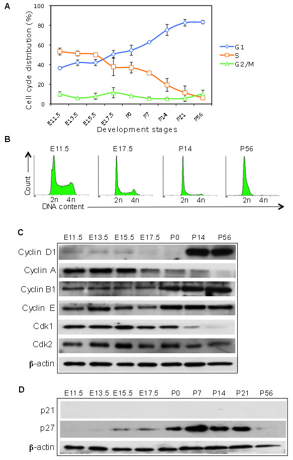
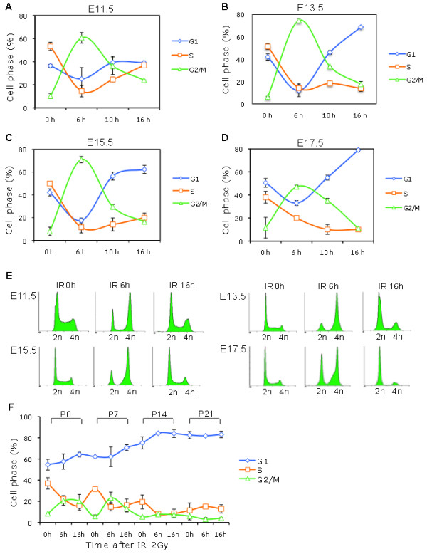
Background: The DNA damage-mediated cell cycle checkpoint is an essential mechanism in the DNA damage response (DDR). During embryonic development, the characteristics of cell cycle and DNA damage checkpoint evolve from an extremely short G1 cell phase and lacking G1 checkpoint to lengthening G1 phase and the establishment of the G1 checkpoint. However, the regulatory mechanisms governing these transitions are not well understood. In this study, pregnant mice were exposed to ionizing radiation (IR) to induce DNA damage at different embryonic stages; the kinetics and mechanisms of the establishment of DNA damage-mediated G1 checkpoint in embryonic liver were investigated.

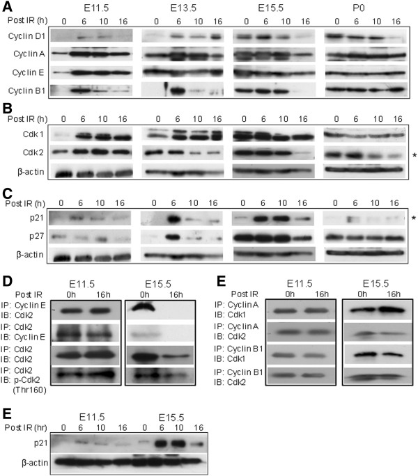
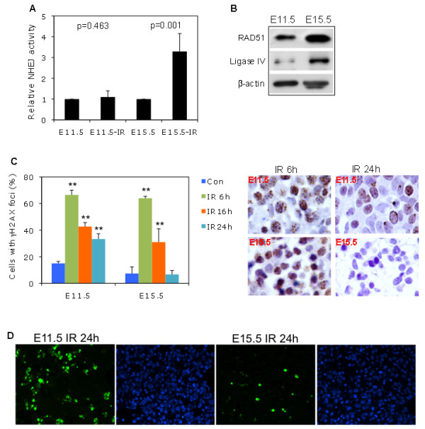
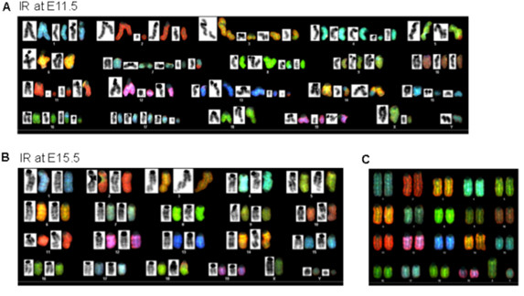
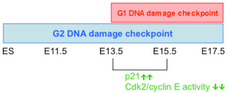
Results: We found that the G2 cell cycle arrest was the first response to DNA damage in early developmental stages. Starting at E13.5/E15.5, IR mediated inhibition of the G1 to S phase transition became evident. Concomitantly, IR induced the robust expression of p21 and suppressed Cdk2/cyclin E activity, which might involve in the initiation of G1 checkpoint. The established G1 cell cycle checkpoint, in combination with an enhanced DNA repair capacity at E15.5, displayed biologically protective effects of repairing DNA double-strand breaks (DSBs) and reducing apoptosis in the short term as well as reducing chromosome deletion and breakage in the long term.

Conclusion: Our study is the first to demonstrate the establishment of the DNA damage-mediated G1 cell cycle checkpoint in liver cells during embryogenesis and its in vivo biological effects during embryonic liver development.


Figures

Figure 1
Figure 1
S and G1 cell cycle distributions in murine liver cells at different stages of development. (A) Single liver cells were isolated from mice at the embryonic stages E11.5, E13.5, E15.5, and E17.5 and at postnatal days 0, 7, 14, 21 and 56, and were then fixed. The DNA content of PI-stained nuclei was determined by flow cytometry. The cells in the G1, S, and G2/M phases were analyzed (Modfit software) and graphed to determine the distribution pattern during murine development. The mean ± SD was from 2 independent experiments. The S-phase cells were dominant at early stages of development. Starting at E17.5, the G1 population started to increase while the S population decreased. (B) Representative histograms of the cell cycle distribution of murine liver tissue at indicated stages. In adult mice (P56), most liver cells were in the G1 cell phase. (C) Liver tissue lysates from mice at the indicated stages of development were prepared for WB analysis of Cdk1, Cdk2 and Cdc25A, and the cyclins D1, E, A, and B1 with β-actin as the loading control. (D) WB analysis of the Cdk inhibitors p21 and p27.
Figure 2
Figure 2
Establishment of the IR-mediated G1 checkpoint in E13.5/E15.5 embryonic liver cells. Pregnant mice at embryonic stages E11.5, E13.5, E15.5, and E17.5 were subjected to 0 or 6 Gy ionizing radiation (IR). At the indicated time points after IR, embryonic liver cells were isolated for PI staining and flow cytometric analysis. The cell cycle arrest patterns were determined at E11.5 (A), E13.5 (B), E15.5 (C), and E17.5 (D) using Modfit software. Each analysis was on a cell pool of 3–5 embryonic livers. The data were the means ± SD from two independent analyses i.e. the embryonic liver cells were from different pregnant mice. Following transient G2 arrest, G1 arrest was observed at E13.5, E15.5, and E17.5 but not at E11.5. (E) Representative histograms of the cell cycle distribution patterns of E11.5, E13.5, E15.5, and E17.5 liver cells after 6 Gy IR at the indicated time points. (F) Mice were subjected to 0 or 2 Gy IR at different postnatal ages. Liver cells were isolated at the indicated time points for cell cycle analysis. Percentages of G1, S, and G2/M cells were determined. The mean ± SD was from 2 independent experiments.
Figure 3
Figure 3
The effects of Cdk2/cyclin E complex down regulation on the G1 checkpoint inE15.5 embryonic liver cells. Pregnant mice were subjected to 0 or 6 Gy IR at E11.5, E13.5, E15.5 and P0. At the indicated time points after IR, embryonic liver cells were isolated for WB. (A) Protein levels of the cyclins D1, A, E, and B1. (B) Protein levels of Cdk1 and Cdk2. In response to IR, Cdk2 expression was down-regulated at E13.5 and E15.5. (C) Protein levels of p21 and p27. In response to IR, a dramatic induction of p21 was observed only at E13.5/E15.5. (D) The lysates from embryonic liver tissues (E11.5 and E15.5) harvested at 0 and 16 hours after IR were immunoprecipitated (IPed) with an antibody against cyclin E, and bound Cdk2 was detected by WB (left panel). The interaction pattern was representative of 2–3 experiments. To confirm the reduced expression of Cdk2/cyclin E complex, the lysates were IPed with an anti-Cdk2 antibody and then immunoblotted (IB) for cyclin E. The same lysates were IPed with anti-Cdk2 antibody and IBed with total Cdk2 and phospho-Cdk2 (Thr 160) (lower panel). (E) The lysates were IPed with antibodies against cyclin A and B1, and bound Cdk1 and Cdk2 were tested by IB. (F) IR-induced p21 expression was significantly enhanced in E15.5 liver cells compared to E11.5 cells.
Figure 4
Figure 4
High DNA damage repair capacity and low apoptosis in E15.5 compared to in E11.5 embryonic liver cells. (A) In vitro NHEJ (non-homologous end-joining) activity was assayed and quantified by plasmid-based quantitative PCR (see Methods). Related NHEJ activity was analyzed by paired Student t test. IR-induced NHEJ activity was significantly higher at E15.5 than at E11.5. (B) Levels of related DNA repair proteins RAD51 and Ligase IV by WB. (C) Left panel: Pregnant mice were exposed to 6 Gy IR. At the indicated time points, E11.5 and E15.5 liver tissue was fixed for γH2AX foci (DSB foci) staining. Nuclear γH2AX foci-positive cells were enumerated. The mean ± SD was from 2 independent experiments. IR induced γH2AX foci-positive cells at different time points were statistically compared to controls, respectively. P value ≤0.05 and ≤ 0.01 was denoted as * and **, respectively. Right panel: Representative γH2AX staining showed that IR-induced DSB foci-positive cells were more dramatically reduced 24 hours after IR in E15.5 than in E11.5 liver cells. (D) Pregnant mice were exposed to 6 Gy IR. At 24 hours after IR, E11.5 and E15.5 liver tissue was fixed for TUNEL staining. Percentages of early apoptotic cells with nuclear FITC labeled nicked DNA vs. DAPI stained liver cells were calculated based on two sections in each case.
Figure 5
Figure 5
Low dose IR at E11.5 and E15.5 and its effects on chromosome abnormality in adult liver cells. (A & B) Pregnant mice were exposed to 0.5 Gy IR at embryonic stages 11.5 or 15.5, and the liver cells were then isolated from the mice at adulthood (7 weeks old) and cultured. Chromosome G-banding and spectral karyotyping (SKY) analysis was performed in metaphase cells. Overall aberrant chromosome at metaphase by G-banding (black color) and SKY (RBG color) in the adult liver cells IR at E11.5 (A) and at E15.5 (B). (C) Normal chromosome at metaphase by SKY.
Figure 6
Figure 6
A proposed model of G1 checkpoint establishment during embryonic liver development. In response to IR damage, G2 cell cycle checkpoint is present in ES cells [8-10] and in E11.5 to E17.5 embryonic liver cells. At E13.5/E15.5 stage, G1 cell cycle checkpoint is established under the regulation of p21-CDK/cyclin E pathway.

Similar articles

Cited by

References

    1. Milyavsky M, Gan OI, Trottier M, Komosa M, Tabach O, Notta F, Lechman E, Hermans KG, Eppert K, Konovalova Z, Omatsky O, Domany E, Meyn MS, Dick JE. A distinctive DNA damage response in human hematopoietic stem cells reveals an apoptosis-independent role for p53 in self-renewal. Cell Stem Cell. 2010;7:186–197. doi: 10.1016/j.stem.2010.05.016. - DOI - PubMed
    1. Fraosina G. The bright and the dark sides of DNA repair in stem cells. J Biomed Biotechnol. 2010;2010:845396. - PMC - PubMed
    1. Poehlmann A, Roessner A. Importance of DNA damage checkpoints in the pathogenesis of human cancers. Pathol Res Pract. 2010;206:591–601. doi: 10.1016/j.prp.2010.06.006. - DOI - PubMed
    1. Orford KW, Scadden DT. Deconstructing stem cell self-renewal: genetic insights into cell cycle regulation. Nat Rev Genet. 2008;9:115–128. doi: 10.1038/nrg2269. - DOI - PubMed
    1. Singh AM, Dalton S. The cell cycle and Myc intersect with mechanisms that regulate pluripotency and reprogramming. Cell Stem Cell. 2009;5:141–149. doi: 10.1016/j.stem.2009.07.003. - DOI - PMC - PubMed

Publication types

MeSH terms