Review
. 2014 May 29:13:129.
doi: 10.1186/1476-4598-13-129.

MUC16 (CA125): tumor biomarker to cancer therapy, a work in progress

Affiliations
Review

MUC16 (CA125): tumor biomarker to cancer therapy, a work in progress

Mildred Felder et al. Mol Cancer. .

Abstract

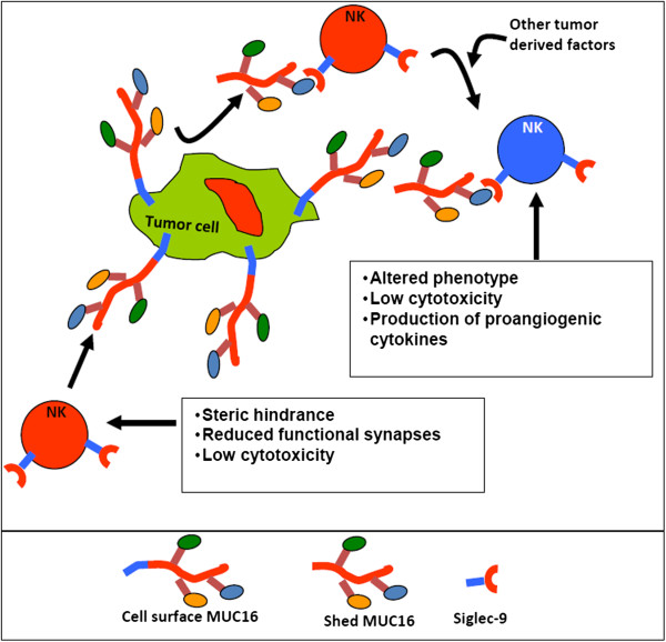
Over three decades have passed since the first report on the expression of CA125 by ovarian tumors. Since that time our understanding of ovarian cancer biology has changed significantly to the point that these tumors are now classified based on molecular phenotype and not purely on histological attributes. However, CA125 continues to be, with the recent exception of HE4, the only clinically reliable diagnostic marker for ovarian cancer. Many large-scale clinical trials have been conducted or are underway to determine potential use of serum CA125 levels as a screening modality or to distinguish between benign and malignant pelvic masses. CA125 is a peptide epitope of a 3-5 million Da mucin, MUC16. Here we provide an in-depth review of the literature to highlight the importance of CA125 as a prognostic and diagnostic marker for ovarian cancer. We focus on the increasing body of literature describing the biological role of MUC16 in the progression and metastasis of ovarian tumors. Finally, we consider previous and on-going efforts to develop therapeutic approaches to eradicate ovarian tumors by targeting MUC16. Even though CA125 is a crucial marker for ovarian cancer, the exact structural definition of this antigen continues to be elusive. The importance of MUC16/CA125 in the diagnosis, progression and therapy of ovarian cancer warrants the need for in-depth research on the biochemistry and biology of this mucin. A renewed focus on MUC16 is likely to culminate in novel and more efficient strategies for the detection and treatment of ovarian cancer.


Figures

Figure 1
Figure 1
MUC16 structure. Model shows the three domains of MUC16 and potential location of the CA125 epitope in a tandem repeat.
Figure 2
Figure 2
OC125 weakly binds to cells that are generally considered negative for MUC16. SKOV-3 cells (identity confirmed by STR analysis) were stained with the primary (1°) antibodies OC125 or VK8 followed by incubation with allophycocyanine (APC)-labeled donkey anti-mouse (DAM) secondary antibody. Binding of the antibodies to SKOV-3 cells was determined on a LSR-II flow cytometer. RT-PCR using MUC16 primers previously reported [178], was used to determine expression of MUC16 in OVCAR-3, ECC-1 and SKOV-3 cells (data not shown).
Figure 3
Figure 3
Cell surface MUC16 is sensitive to proteolysis. A, Binding of OC125 and VK8 to OVCAR-3 cells that were harvested with trypsin or EDTA containing media was determined by flow cytometry. A significant decrease in VK8 binding to cells harvested after trypsin treatment was observed whereas under these conditions, OC125 binding was less affected. B, Reduction of the trypsinized OVCAR-3 cells with dithiothreitol (DTT) or 2-mercaptoethanol (2-ME) did not result in additional loss of OC125 binding to the cells. In all experiments, a fluorescently tagged goat anti-mouse antibody was used for detection.
Figure 4
Figure 4
MUC16 on the surface of ovarian cancer cells. OVCAR-3 cells were labeled with VK8 followed by colloidal gold nanoparticles conjugated with goat anti-mouse secondary antibody. A, low magnification secondary electron image of two labeled OVCAR-3 cells. B, Scanning Electron Microscopy (SEM) image of OVCAR-3 showing colloidal gold nanoparticles binding to cell surface and microvilli. C, Back scattered electron image of same cell surface shown in B, clearly showing the colloidal gold nanoparticles. Bright spots (some indicated by bright arrows) in B and C are the colloidal gold nanoparticles. OVCAR-3 cells are not labeled with colloidal gold nanoparticles in the absence of VK-8 (data not shown).
Figure 5
Figure 5
Model for MUC16-induced NK cell inhibition. MUC16 released from tumors binds to naïve NK cells (shown in red) and along with other tumor derived factors induces a phenotypic and functional change. The altered NK cells (shown in blue) secrete cytokines that promote angiogenesis. The cell surface bound MUC16, on the other hand, acts as an anti-adhesive mucin and blocks the interaction between the NK cells and ovarian tumor cells thereby preventing cancer cell cytolysis.
Figure 6
Figure 6
Analysis of data on MUC16 available through TCGA analysis of ovarian cancer samples. A TCGA data set [148] was analyzed to determine differential expression of prominent cancer-related genes between normal ovarian tissue and ovarian carcinoma (heat map in top panel). Although MUC16 is upregulated in the cancer samples (expression in cancer specimens is double than in normal) this difference is not significant (p-value = 0.1), probably because there were only 8 normal samples in TCGA dataset. MUC16 is compared to other prominent mutated genes reported in the original TCGA report [148]. Of these genes, only BRCA2 is differentially expressed between normal and cancer (p-value <0.001) samples. Analysis of mutated and wild-type MUC16 expression in samples from ovarian cancer patients listed in TCGA is shown in the heat map in the lower panel. Mutated MUC16 is also mildly over-expressed as compared to wild-type mucin (expression Wild-type/Mutated = 0.94), however this difference is not significant (p-value = 0.84). MUC16 is compared to other genes reported to be highly mutated at the original report of TCGA. None of the other genes is differentially expressed between MUC16-wild-type and MUC16-mutated ovarian cancer specimens, indicating no association between MUC16 mutation status and key genes expression in this dataset (p-value <0.05). B, Survival of ovarian cancer patients with wild-type and mutated MUC16 was compared. Although a trend was observed suggesting worse outcome in patients with mutated MUC16, the difference was not statistically significant (p-value = 0.28). C, MUC16 (both mutated and wild-type) expression was divided into quartiles. Survival of patients in each of these quartiles was not significantly different.

Similar articles

Cited by

References

    1. Duffy MJ, Bonfrer JM, Kulpa J, Rustin GJ, Soletormos G, Torre GC, Tuxen MK, Zwirner M. CA125 in ovarian cancer: European group on tumor markers guidelines for clinical use. Int J Gynecol Cancer. 2005;15:679–691. doi: 10.1111/j.1525-1438.2005.00130.x. - DOI - PubMed
    1. Sturgeon CM, Duffy MJ, Stenman UH, Lilja H, Brunner N, Chan DW, Babaian R, Bast RC Jr, Dowell B, Esteva FJ, Haglund C, Harbeck N, Hayes DF, Holten-Andersen M, Klee GG, Lamerz R, Looijenga LH, Molina R, Nielsen HJ, Rittenhouse H, Semjonow A, Shih Ie M, Sibley P, Soletormos G, Stephan C, Sokoll L, Hoffman BR, Diamandis EP. National Academy of Clinical Biochemistry laboratory medicine practice guidelines for use of tumor markers in testicular, prostate, colorectal, breast, and ovarian cancers. Clin Chem. 2008;54:e11–e79. doi: 10.1373/clinchem.2008.105601. - DOI - PubMed
    1. Seltzer V, Drukker BH, Gillespie BW, Gossfeld LM, Grigsby PW, Harvey HA, Hendricks CB, Hummel S, Makuch RW, Monaco GP, Parham GP, Sawyers CL, West RJ, Alberts DS, Anderson B, Averette HE, Bast RC, Christian MC, Colombo N, Creasman WT, John P, Curtin JP, Gershenson DM, Hoskins WJ, Karlan BY, Kramer BS, Markman M, McGuire WP, Ozols RF, Pecorelli S. et al.NIH consensus conference: ovarian cancer: screening, treatment, and follow-up. NIH consensus development panel on ovarian cancer. JAMA. 1995;273:491–497. doi: 10.1001/jama.1995.03520300065039. - DOI - PubMed
    1. Rustin GJ, Quinn M, Thigpen T, du Bois A, Pujade-Lauraine E, Jakobsen A, Eisenhauer E, Sagae S, Greven K, Vergote I, Cervantes A, Vermorken J. Re: new guidelines to evaluate the response to treatment in solid tumors (ovarian cancer) J Natl Cancer Inst. 2004;96:487–488. - PubMed
    1. Vasudev NS, Trigonis I, Cairns DA, Hall GD, Jackson DP, Broadhead T, Buxton J, Hutson R, Nugent D, Perren TJ. The prognostic and predictive value of CA-125 regression during neoadjuvant chemotherapy for advanced ovarian or primary peritoneal carcinoma. Arch Gynecol Obstet. 2011;284:221–227. doi: 10.1007/s00404-010-1655-2. - DOI - PMC - PubMed

Publication types

MeSH terms