

Histopathological evaluation of non-melanoma skin cancer
- PMID: 24886534
- PMCID: PMC4046093
- DOI: 10.1186/1477-7819-12-159
Histopathological evaluation of non-melanoma skin cancer
Abstract
Background: Non-melanoma skin cancers (NMSCs) are the most frequently seen cancers worldwide.
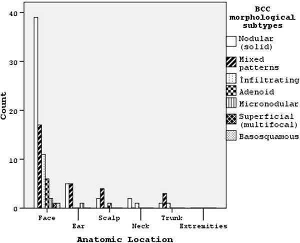
Methods: The medical records of patients diagnosed with basal cell carcinoma (BCC) and squamous cell carcinoma (SCC) in Hatay Antakya pathology laboratory between January 2010 and September 2012 were retrospectively included in the study. Tumors were categorized according to age, gender, anatomical localization, type, solitary-multiplicity, tumor diameter (0 to 2 mm, 2.1 to 6 mm and >6.1 mm), and presence of ulceration (BCCs), and morphological subtype, histopatological features and grades (SCCs).
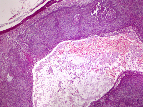
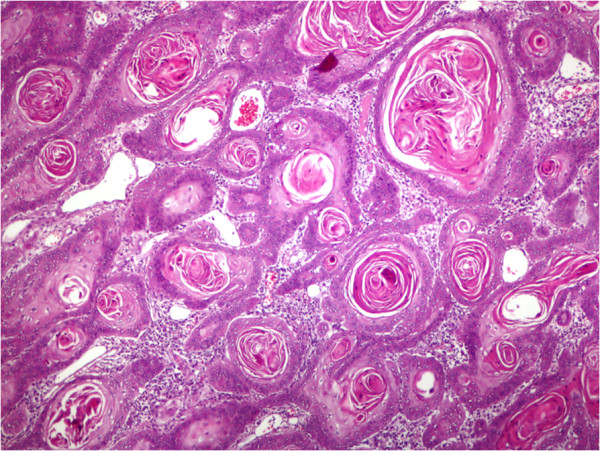
Results: A total of 136 tumors in 127 NMSC cases were examined. Solitary tumors were seen in 118 (92.9%), and multiple tumors in 9 (7.1%) patients. Mean age of the patients was 68.5 ± 13 years. BCC was observed in 96 (75.6%) and SCC in 31 (24.4%) patients. Mean diameter of all types of solitary and multiple tumors was 7.42 ± 3.49 mm. Nodular subtype focal cystic changes were observed in 49 (47.6%) patients. All tumors (solitary and multiple) were seen on the face (67.6%), scalp (11.8%), and ear (11%). Well differentiated SCCs were detected in 20 cases (64.5%); ulceration was observed in 58.1% of all tumors.
Conclusions: Epidemiologic and histopathological investigations, routine skin scanning performed on the elderly population and dermatological examination will help to improve efficient health applications.
Figures
Similar articles
-
Clinicopathological Pattern of Nonmelanoma Skin Cancer in Kuwait: A Retrospective Study.Med Princ Pract. 2024;33(2):133-138. doi: 10.1159/000536010. Epub 2023 Dec 29. Med Princ Pract. 2024. PMID: 38160671 Free PMC article.
-
Non-melanoma skin cancers between the years of 1990 and 1999 in Izmir, Turkey: demographic and clinicopathological characteristics.J Dermatol. 2003 Feb;30(2):123-31. doi: 10.1111/j.1346-8138.2003.tb00359.x. J Dermatol. 2003. PMID: 12692379
-
Non-melanoma Skin Cancer - a Clinicopathological Study of Patients with Basal Cell Carcinoma and Squamous Cell Carcinoma.Klin Onkol. 2017 Winter;31(1):40-45. doi: 10.14735/amko201840. Klin Onkol. 2017. PMID: 29488777 English.
-
UV-Induced Molecular Signaling Differences in Melanoma and Non-melanoma Skin Cancer.Adv Exp Med Biol. 2017;996:27-40. doi: 10.1007/978-3-319-56017-5_3. Adv Exp Med Biol. 2017. PMID: 29124688 Review.
-
Non-melanoma skin cancer: what drives tumor development and progression?Carcinogenesis. 2005 Oct;26(10):1657-67. doi: 10.1093/carcin/bgi123. Epub 2005 May 19. Carcinogenesis. 2005. PMID: 15905207 Review.
Cited by
-
Analysis of the occurrence and distribution of primary and recurrent basal cell carcinoma of head and neck coupled to the assessment of tumor microenvironment and Sonic hedgehog signaling.Rom J Morphol Embryol. 2020 Jul-Sep;61(3):821-831. doi: 10.47162/RJME.61.3.20. Rom J Morphol Embryol. 2020. PMID: 33817723 Free PMC article.
-
Lack of efficacy of imiquimod in patients with basal cell carcinoma previously treated with rituximab for B cell lymphoma: two case reports.J Med Case Rep. 2016 Mar 11;10:57. doi: 10.1186/s13256-016-0834-6. J Med Case Rep. 2016. PMID: 26968156 Free PMC article.
-
Diet phytochemicals and cutaneous carcinoma chemoprevention: A review.Pharmacol Res. 2017 May;119:327-346. doi: 10.1016/j.phrs.2017.02.021. Epub 2017 Feb 24. Pharmacol Res. 2017. PMID: 28242334 Free PMC article. Review.
-
TP53 Gene Polymorphisms and Occupational Skin Cancer Risks for Workers of Glass Fiber Manufacture.Iran J Public Health. 2017 Nov;46(11):1495-1501. Iran J Public Health. 2017. PMID: 29167767 Free PMC article.
-
Rare association of cystic squamous cell carcinoma and small lymphocytic B cell lymphoma: successful surgical approach.Wien Med Wochenschr. 2017 Apr;167(5-6):104-109. doi: 10.1007/s10354-016-0510-x. Epub 2016 Sep 8. Wien Med Wochenschr. 2017. PMID: 27631871
References
-
- Weedon D. In: Weedon’s Skin Pathology. 3. Weedon D, editor. London: Churchill Livingstone; 2010. Tumors of the epidermis; pp. 668–703.
-
- Weedon D, Marks R, Kao GF, Harword CA. In: World Health Organization Classification of Tumours. Pathology & Genetics of Skin Tumours. LeBoit PE, Burg G, Weedon D, Sarasain A, editor. Lyon: IARC Press; 2006. Keratinocytic Tumours; pp. 9–47.
-
- Rao P, Liegeois NJ, McNiff JM, Nghiem P, Prieto VG, Smith MT, Smoller BR, Wick MR, Frishberg D. Protocol for the Examination of Specimens From Patients With Squamous Cell Carcinoma of the Skin. Washington: College of American Pathologists (CAP); pp. 1996–2010. http://www.cap.org (accessed 13 September 2012)
Publication types
MeSH terms
LinkOut - more resources
Full Text Sources
Other Literature Sources
Medical
Research Materials
