. 2014 May 22:10:31.
doi: 10.1186/1744-8069-10-31.

Artemin growth factor increases nicotinic cholinergic receptor subunit expression and activity in nociceptive sensory neurons

Affiliations

Artemin growth factor increases nicotinic cholinergic receptor subunit expression and activity in nociceptive sensory neurons

Kathryn M Albers et al. Mol Pain. .

Abstract

Background: Artemin (Artn), a member of the glial cell line-derived growth factor (GDNF) family, supports the development and function of a subpopulation of peptidergic, TRPV1-positive sensory neurons. Artn (enovin, neublastin) is elevated in inflamed tissue and its injection in skin causes transient thermal hyperalgesia. A genome wide expression analysis of trigeminal ganglia of mice that overexpress Artn in the skin (ART-OE mice) showed elevation in nicotinic acetylcholine receptor (nAChR) subunits, suggesting these ion channels contribute to Artn-induced sensitivity. Here we have used gene expression, immunolabeling, patch clamp electrophysiology and behavioral testing assays to investigate the link between Artn, nicotinic subunit expression and thermal hypersensitivity.

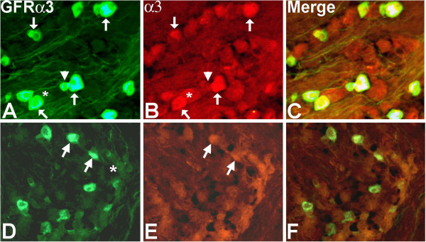
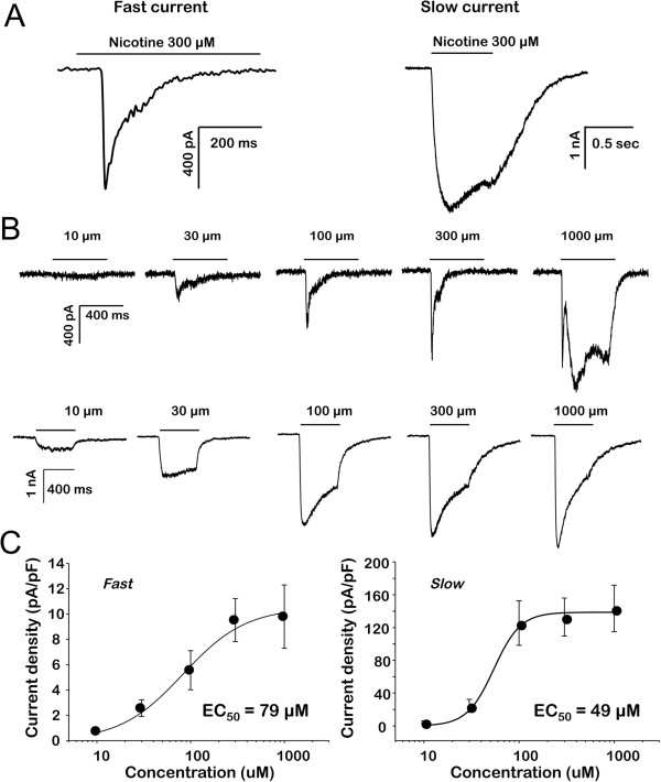
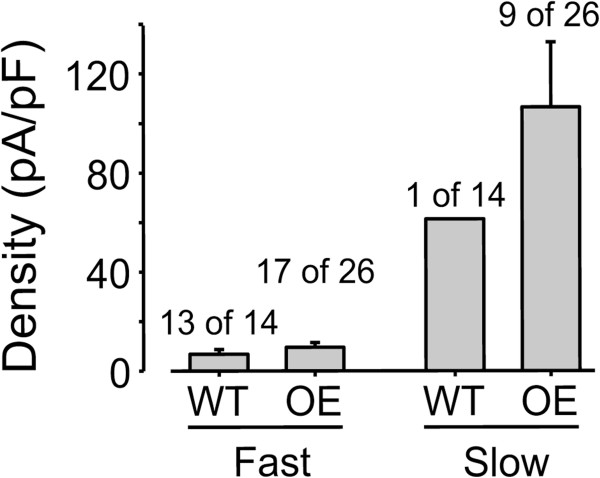
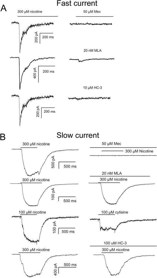
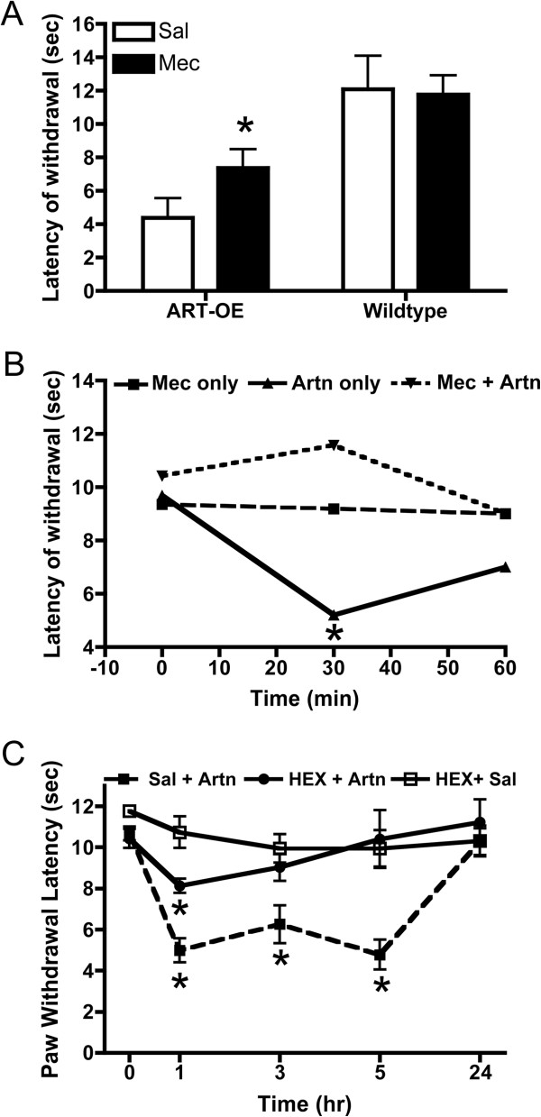
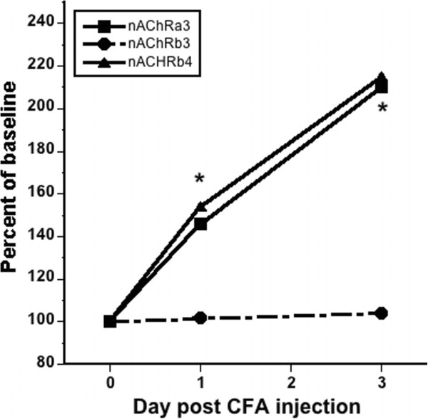
Results: Reverse transcriptase-PCR validation showed increased levels of mRNAs encoding the nAChR subunits α3 (13.3-fold), β3 (4-fold) and β4 (7.7-fold) in trigeminal ganglia and α3 (4-fold) and β4 (2.8-fold) in dorsal root ganglia (DRG) of ART-OE mice. Sensory ganglia of ART-OE mice had increased immunoreactivity for nAChRα3 and exhibited increased overlap in labeling with GFRα3-positive neurons. Patch clamp analysis of back-labeled cutaneous afferents showed that while the majority of nicotine-evoked currents in DRG neurons had biophysical and pharmacological properties of α7-subunit containing nAChRs, the Artn-induced increase in α3 and β4 subunits resulted in functional channels. Behavioral analysis of ART-OE and wildtype mice showed that Artn-induced thermal hyperalgesia can be blocked by mecamylamine or hexamethonium. Complete Freund's adjuvant (CFA) inflammation of paw skin, which causes an increase in Artn in the skin, also increased the level of nAChR mRNAs in DRG. Finally, the increase in nAChRs transcription was not dependent on the Artn-induced increase in TRPV1 or TRPA1 in ART-OE mice since nAChRs were elevated in ganglia of TRPV1/TRPA1 double knockout mice.

Conclusions: These findings suggest that Artn regulates the expression and composition of nAChRs in GFRα3 nociceptors and that these changes contribute to the thermal hypersensitivity that develops in response to Artn injection and perhaps to inflammation.


Figures

Figure 1
Figure 1
Immunolabeling shows nAChRα3 is expressed in GFRα3-neurons. Labeling of nAChRα3-positive neurons in ART-OE ganglia (A-C) is more intense compared to WT ganglia (D-F). Quantitative analysis showed the percentage of GFRα3 neurons that express nAChRα3 is greater in ganglia of ART-OE mice (46%) compared to WT mice (9.7%) (p < 0.05%, Student’s t test). Arrows indicate GFRα3/nAChRα3 positive neurons; asterisks mark nAChRα3 neurons that are not GFRα3 positive; arrowhead shows GFRα3 neuron that is nAChRα3 negative.
Figure 2
Figure 2
Nicotine evoked currents in acutely dissociated DRG neurons. Nicotine was applied for 500 ms to neurons held at −60 mV via a fast application system. A. Two types of currents were detected in mouse DRG neurons that were distinguishable based on differences in activation and inactivation kinetics: A fast current (left) that activated and inactivated rapidly and a slow current (right) that activated and inactivated more slowly. B. Fast (top traces) and slow (bottom traces) currents were activated by nicotine over a comparable concentration range. Fast and slow currents were evoked from the same neuron in response to increasing concentrations of nicotine. A third even more slowly activating current (top trace, right side) was evoked at higher (1000 μM) concentrations of nicotine and was present in most neurons with fast current as well as a significant number of neurons without either fast or slow current. C. Concentration response data from individual neurons with fast (n = 26) or slow (n = 16) current were pooled and fitted with a Hill equation to estimate the maximal evoked current and the concentration at which a current 50% of maximal was evoked (EC50).
Figure 3
Figure 3
Nicotine-evoked current density. Current density was determined by dividing the peak-evoked current in response to 300 μM nicotine by the membrane capacitance. Pooled data from neurons with fast and slow current indicate that the slow current density is significantly greater than that of the fast current. The proportion of neurons from wild type (WT) and ART-OE (OE) mice with evoked current that was either fast or slow is indicated above each bar.
Figure 4
Figure 4
Pharmacological characterization of nicotine evoked currents. A. The fast current was blocked by the non-specific nicotinic receptor antagonist mecamylamine (Mec, top trace), the α7-subunit antagonist MLA (middle trace) and the TRPA1 “selective” antagonist HC-30031 (HC, bottom traces). Currents are from three different neurons in response to nicotine applied with a 5 min inter-stimulus interval before and after the application of antagonist. B. The slow current was also blocked by Mec (top row) but was resistant to MLA (second row) and activated by the α4β3 agonist cytisine (third row). The slow current was also resistant to HC-30031. As in A, current in each row was evoked from a different neuron. All data in A and B were from ART-OE mice, but comparable data for the fast current were obtained from WT mice.
Figure 5
Figure 5
nAChR antagonists block Artn-induced heat hyperalgesia. A. WT (n = 4) and ART-OE (n = 6) mice were injected with either 10 μl saline (right foot) or 10 μl Mec (left foot, 1 mg/kg) and latencies of withdrawal measured for each foot 60 min post injection. As previously reported [10], ART-OE mice exhibit lower heat thresholds compared with WT mice (compare saline treated groups). Mec partially blocks heat hyperalgesia in ART-OE mice (p < 0.05, ANOVA) and did not affect thermal sensitivity in WT mice. B. Footpads of WT mice were injected with 1 mg/kg Mec or saline 30 min prior to injection of Artn (200 ng) or saline. Mec injection had no effect on thermal sensitivity. Artn injection alone increased heat hyperalgesia whereas Mec + Artn blocked hyperalgesia. C. Mice (n = 6) were injected in the left footpad with saline (10 μl) 30 min prior to Artn injection (200 ng). Other mice (n = 6) were injected with HEX (10 μl, 1 mg/kg) followed by Artn. A third set (n = 6) were injected with HEX followed by saline. HEX alone did not affect thermal sensitivity. HEX blocked thermal sensitivity caused by Artn at 1 h, 3 h and 5 h post injection. Measures were made in a blinded manner. Asterisks indicate p < 0.05.
Figure 6
Figure 6
CFA-induced skin inflammation increases nAChR mRNA levels in DRG. The percent of baseline expression of nAChR mRNAs in lumbar DRG of uninjected mice (n = 5) and CFA injected mice (n = 6) was compared using SYBR green qRT-PCR at 1 d and 3 d post injection of CFA into footpads of C57BL/6 J mice. Percent change normalized to gapdh is plotted. Asterisks indicate significant change in β4 at day 1 and α3 at day 3 (two-way ANOVA with Bonferroni posttests with p < 0.05).

Similar articles

Cited by

References

    1. Gold MS, Gebhart GF. Nociceptor sensitization in pain pathogenesis. Nat Med. 2010;16:1248–1257. doi: 10.1038/nm.2235. - DOI - PMC - PubMed
    1. Forrest SL, Keast JR. Expression of receptors for glial cell line-derived neurotrophic factor family ligands in sacral spinal cord reveals separate targets of pelvic afferent fibers. J Comp Neurol. 2008;506:989–1002. doi: 10.1002/cne.21535. - DOI - PMC - PubMed
    1. Orozco OE, Walus L, Sah DW, Pepinsky RB, Sanicola M. GFRalpha3 is expressed predominantly in nociceptive sensory neurons. Eur J Neurosci. 2001;13:2177–2182. doi: 10.1046/j.0953-816x.2001.01596.x. - DOI - PubMed
    1. Weinkauf B, Rukwied R, Quiding H, Dahllund L, Johansson P, Schmelz M. Local gene expression changes after UV-irradiation of human skin. PLoS One. 2012;7:e39411. doi: 10.1371/journal.pone.0039411. - DOI - PMC - PubMed
    1. Murota H, Izumi M, Abd El-Latif MI, Nishioka M, Terao M, Tani M, Matsui S, Sano S, Katayama I. Artemin causes hypersensitivity to warm sensation, mimicking warmth-provoked pruritus in atopic dermatitis. J Allergy Clin Immunol. 2012;130:671–682. doi: 10.1016/j.jaci.2012.05.027. e674. - DOI - PubMed

Publication types

MeSH terms

LinkOut - more resources