. 2014 May 21:11:38.
doi: 10.1186/1742-4690-11-38.

Switching and emergence of CTL epitopes in HIV-1 infection

Affiliations

Switching and emergence of CTL epitopes in HIV-1 infection

Chungyong Han et al. Retrovirology. .

Abstract

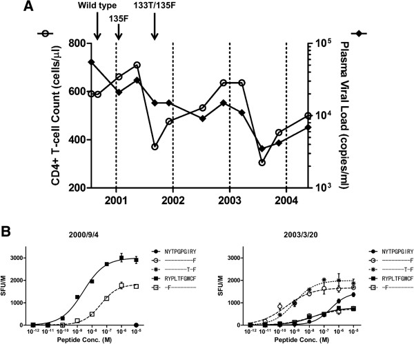
Background: Human Leukocyte Antigen (HLA) class I restricted Cytotoxic T Lymphocytes (CTLs) exert substantial evolutionary pressure on HIV-1, as evidenced by the reproducible selection of HLA-restricted immune escape mutations in the viral genome. An escape mutation from tyrosine to phenylalanine at the 135th amino acid (Y135F) of the HIV-1 nef gene is frequently observed in patients with HLA-A*24:02, an HLA Class I allele expressed in ~70% of Japanese persons. The selection of CTL escape mutations could theoretically result in the de novo creation of novel epitopes, however, the extent to which such dynamic "CTL epitope switching" occurs in HIV-1 remains incompletely known.

Results: Two overlapping epitopes in HIV-1 nef, Nef126-10 and Nef134-10, elicit the most frequent CTL responses restricted by HLA-A*24:02. Thirty-five of 46 (76%) HLA-A*24:02-positive patients harbored the Y135F mutation in their plasma HIV-1 RNA. Nef codon 135 plays a crucial role in both epitopes, as it represents the C-terminal anchor for Nef126-10 and the N-terminal anchor for Nef134-10. While the majority of patients with 135F exhibited CTL responses to Nef126-10, none harboring the "wild-type" (global HIV-1 subtype B consensus) Y135 did so, suggesting that Nef126-10 is not efficiently presented in persons harboring Y135. Consistent with this, peptide binding and limiting dilution experiments confirmed F, but not Y, as a suitable C-terminal anchor for HLA-A*24:02. Moreover, experiments utilizing antigen specific CTL clones to recognize endogenously-expressed peptides with or without Y135F indicated that this mutation disrupted the antigen expression of Nef134-10. Critically, the selection of Y135F also launched the expression of Nef126-10, indicating that the latter epitope is created as a result of escape within the former.

Conclusions: Our data represent the first example of the de novo creation of a novel overlapping CTL epitope as a direct result of HLA-driven immune escape in a neighboring epitope. The robust targeting of Nef126-10 following transmission (or in vivo selection) of HIV-1 containing Y135F may explain in part the previously reported stable plasma viral loads over time in the Japanese population, despite the high prevalence of both HLA-A*24:02 and Nef-Y135F in circulating HIV-1 sequences.


Figures

Figure 1
Figure 1
The association between the two overlapping epitopes and the Y135F mutation. (A) HIV-1 map of representative proteins and the locations of HLA-A*24:02-restricted epitopes used in this study are shown (arrow). The positional relation and the sequence information around Nef126-10 and Nef134-10 epitopes are also shown. (B) Immune responses to 11 epitopes were assessed by IFN-γ ELISpot assay by using expanded PBMCs from 46 HIV-1 patients. Epitope names below the chart indicate peptides used in the assay; [Protein][Location(HXB2 numbering)]-[Amino acid length]. Each point represents the average SFU of duplicate wells after subtraction of background in each individual. Each stick represents response rate in 46 individuals. (C) The Nef126-10/Nef134-10-specific response rates among individuals harboring either Nef135Y or Nef135F were assessed. Black bars indicate number of responders to each epitope, and white bars for non-responders.
Figure 2
Figure 2
Epitope characterization. (A) Binding affinity of each peptide to HLA-A*24:02 was tested by using T2-A24 cell line. All peptides except negative control peptide, VPLDKDFRKY, which might not bind to HLA-A*24:02, show HLA-A*24:02 binding in the peptide concentration of 10-4 M. Each point and bar represents the average and the standard deviation of normalized MFI from three independent experiments. (B) 293FT-A24DRm-CY0 cells were pulsed with serially diluted peptides and co-cultured with CTL clones specific to Nef134-10 (H27-9) or Nef126-10 (I30-1). Epitope recognition was determined by normalizing quantity of IFN-γ secretion. For normalization, all values were divided by IFN-γ secretion with wild type peptide concentration of 9 μM. Neither clone showed specificity to the negative control, Gag28-9 peptide (KYKLKHIVW) a binder of HLA-A*24:02. Each point and bar show average and standard deviation of three independent experiments.
Figure 3
Figure 3
Recognition of endogenously derived epitope. (A) Vector construction. The vector expresses EGFP as a transfection marker and a fusion protein that consists of mini-Nef gene coding 31 amino acids of Nef123-Nef153 region and GlyGlyGlyGlySer linker and renilla luciferase. The 133I/135F and 133T/135F mutant vectors as well as wild type vector were constructed. Mini-Nef sequence that corresponds to the actual Nef gene is shown. (B) By measuring luciferase activity, quantity of the generated mini-Nef was assessed. No significant differences in mini-Nef generation between wild type, 135F and 133T/135F were observed. Each stick and bar indicates average and standard deviation of three independent experiments. (C) The vector transfected cells were incubated with either H27-9 or I30-1, and IFN-γ secretion was quantified by ELISA and normalized to determine the epitope recognition. For normalization, each value was divided by IFN-γ secretion with wild type peptide concentration of 9 μM. Mock as well as mGag, transfected with mGag(wt)-hRluc-EGFP, were measured as negative controls. Each stick and bar indicates average and standard deviation of three independent experiments. The significance was calculated by unpaired Student’s t-test; **, p < 0.01; ***, p < 0.001.
Figure 4
Figure 4
Longitudinal analysis in one individual. (A) Time courses of the surrogate markers are shown. CD4+ T cell count (left axis, white circle) and pVL (right axis, black circle) are plotted on the chart. Each arrow indicates the earliest time point when the sequence above the arrow was observed. (B) IFN-γ ELISpot assay was performed by using serially diluted peptide set and expanded PBMCs. Two time points were tested for the assay. At the earlier point, 2000/9/4, the patient harbored wild type sequence around the Nef126-10/Nef134-10 region, and at the latter point, 2003/3/20, the sequence displayed 133T/135F. Mutants as well as wild type peptides of Nef126-10 or Nef134-10 epitopes were used in the assay. Each point represents the average SFU of duplicate wells after subtraction of background, and each error bar shows standard deviation.
Figure 5
Figure 5
Relevance of I133T mutation to Y135F and its impact on CTL responses. (A) The cross-sectional analysis of the co-variation relationship between Nef codons 133 and 135. Nef codons containing gaps or amino acid mixtures were excluded from analysis, leaving 941 and 924 sequences for analysis of codon pairs 135F/133T and 135Y/133I, respectively. (B) Kaplan-Meier plot of the emergence of Y135F and I133T mutations in a multicenter longitudinal acute/early infection cohort comprising 16 HLA-A*24:02-expressing persons. (C) Within 24 patients who harboring 133I/135F or 133T/135F mutant, correlation between the magnitude of peptide-specific response and pVL was examined. Peptide-specific response was determined by using ex vivo IFN-γ ELISpot assay with frozen PBMCs. Each point is plotted according to the average SFU of duplicate wells and the measured pVL at the sampling point. Statistical significance of the correlation between the magnitude and pVL was calculated by Spearman’s rank correlation.
Figure 6
Figure 6
Impact of I133T mutation on the structures of pHLA and responding T cell subsets. (A) Top; Side view of structures of the Nef126-10(8I10F) (colored purple) and Nef126-10(8T10F) (colored green) is shown. Superimposition is based on the HLA-A*24:02 peptide binding domain. Water molecules are represented in blue and yellow spheres for the Nef126-10(8I10F) and Nef126-10(8T10F) epitopes, respectively. The number of peptide residues is labeled in black through P1 to P10. The HLA-A*24:02 is represented as a dark grey (Nef126-10(8T10F) and light grey cartoon. (B) The presence of the responses to each peptide was examined by ex vivo ELISpot assay. A pair of sticks, Nef126-10(8I10F) (purple) and Nef126-10(8T10F) (green), represents the average SFU of duplicate wells to each peptide in one patient. Four individuals on the left side harbored 133I/135F mutant and 10 individuals on the right side harbored 133T/135F mutant. (C) Functional avidity curve was drawn for 9 subjects who harbored Nef133T/135F sequence. From the curve, SD50 was calculated and projected in this chart. Nef126-10(8I10F)-specific CTL responses (purple) showed significantly higher functional avidity compared to Nef126-10(8T10F) (green). Each point represents functional avidity for specific peptide in one individual. The significance between two groups was calculated by Mann–Whitney U-test; *, p < 0.05.

References

    1. McMichael AJ, Rowland-Jones SL. Cellular immune responses to HIV. Nature. 2001;410(6831):980–987. doi: 10.1038/35073658. - DOI - PubMed
    1. Walker BD, Burton DR. Toward an AIDS vaccine. Science. 2008;320(5877):760–764. doi: 10.1126/science.1152622. - DOI - PubMed
    1. Kawana A, Tomiyama H, Takiguchi M, Shioda T, Nakamura T, Iwamoto A. Accumulation of specific amino acid substitutions in HLA-B35-restricted human immunodeficiency virus type 1 cytotoxic T lymphocyte epitopes. AIDS Res Hum Retrovir. 1999;15(12):1099–1107. doi: 10.1089/088922299310395. - DOI - PubMed
    1. Furutsuki T, Hosoya N, Kawana-Tachikawa A, Tomizawa M, Odawara T, Goto M, Kitamura Y, Nakamura T, Kelleher AD, Cooper DA, Iwamoto A. Frequent transmission of cytotoxic-T-lymphocyte escape mutants of human immunodeficiency virus type 1 in the highly HLA-A24-positive Japanese population. J Virol. 2004;78(16):8437–8445. doi: 10.1128/JVI.78.16.8437-8445.2004. - DOI - PMC - PubMed
    1. Phillips RE, Rowland-Jones S, Nixon DF, Gotch FM, Edwards JP, Ogunlesi AO, Elvin JG, Rothbard JA, Bangham CR, Rizza CR, McMichael A. Human immunodeficiency virus genetic variation that can escape cytotoxic T cell recognition. Nature. 1991;354(6353):453–459. doi: 10.1038/354453a0. - DOI - PubMed

Publication types

MeSH terms

Substances

Associated data