

Low-dose insulin-like growth factor binding proteins 1 and 2 and angiopoietin-like protein 3 coordinately stimulate ex vivo expansion of human umbilical cord blood hematopoietic stem cells as assayed in NOD/SCID gamma null mice
- PMID: 24886724
- PMCID: PMC4076633
- DOI: 10.1186/scrt460
Low-dose insulin-like growth factor binding proteins 1 and 2 and angiopoietin-like protein 3 coordinately stimulate ex vivo expansion of human umbilical cord blood hematopoietic stem cells as assayed in NOD/SCID gamma null mice
Abstract
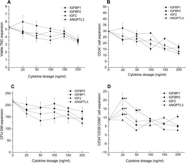
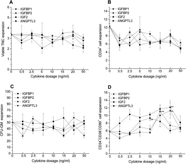
Introduction: Insulin-like growth factors (IGFs), IGF binding proteins (IGFBPs) and angiopoietin-like proteins (ANGPTLs) can enhance the ex vivo expansion of hematopoietic stem cells (HSCs) when used with a standard cytokine cocktail of stem cell factor (SCF), thrombopoietin (TPO) and FLT3 ligand (FL). In order to determine the optimal dose and combination of IGFs, IGFBPs and ANGPTLs, serial dilution and full permutation of IGFBP1, IGFBP2, IGF2 and ANGPTL3 were applied on a cryopreserved umbilical cord blood mononuclear cell (UCB-MNC) ex vivo expansion system.
Methods: In this system, 4 × 10⁵ cells/ml of UCB-MNCs were inoculated in serum-free Stemspan® medium (Stemcell technologies, vancouver, BC, Canada) supplied with standard basal cytokine combination of 100 ng/ml SCF, 50 ng/ml FL and 100 ng/ml TPO and supported by a bone marrow mesenchymal stromal cell layer.
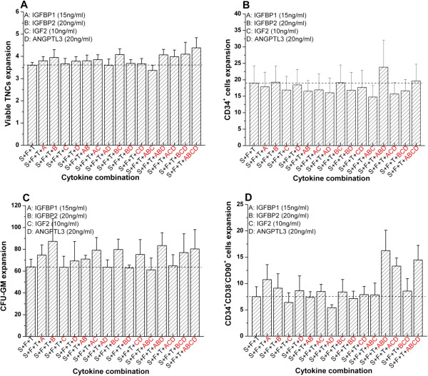
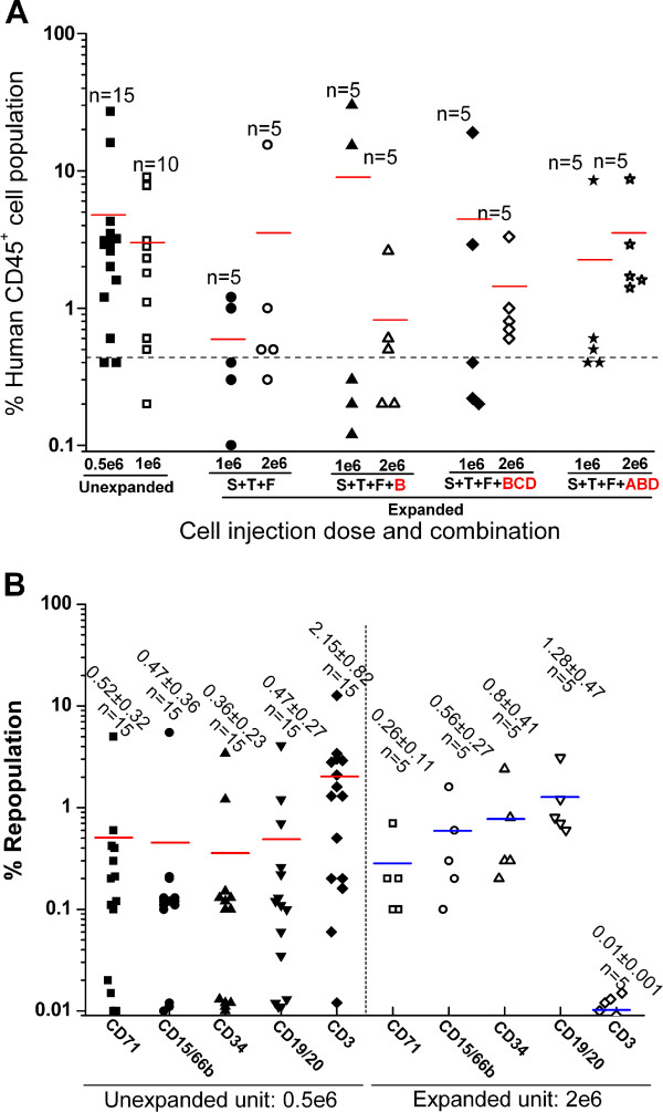
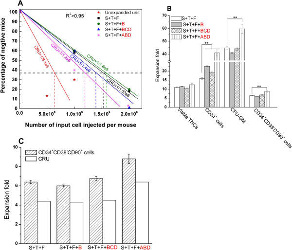
Results: Paradoxically, experiment results showed that the highest expansion of CD34+CD38-CD90+ primitive progenitor was stimulated by cytokine combination of SCF + TPO + FL + IGFBP1 + IGFBP2 + ANGPTL3 at a low dose of 15 ng/ml IGFBP1 and 20 ng/ml IGFBP2 and ANGPTL3. This ex vivo expansion was further validated in 8-week-old to 10-week-old nonobese diabetic/severe combined immunodeficiency interleukin 2 gamma chain null (NOD/SCID-IL2Rγ-/-) mice. Limiting dilution assay showed excellent correlation between the HSC ex vivo surface marker of CD34+CD38-CD90+ and the in vivo competitive repopulating unit (CRU) functional assay.
Conclusion: IGFBP1, IGFBP2, IGF2 and ANGPTL3 can stimulate the expansion of CD34+CD38-CD90+ primitive progenitor at low dose. The optimal combination comprises IGFBP1, IGFBP2 and ANGPTL3 together with the standard cytokine cocktail of SCF, FL and TPO. The CD34+CD38-CD90+ phenotype can serve as a surrogate ex vivo surface marker for HSCs due to consistency with the in vivo CRU functional assay.
Figures
Similar articles
-
Angiopoietin-like 5 and IGFBP2 stimulate ex vivo expansion of human cord blood hematopoietic stem cells as assayed by NOD/SCID transplantation.Blood. 2008 Apr 1;111(7):3415-23. doi: 10.1182/blood-2007-11-122119. Epub 2008 Jan 17. Blood. 2008. PMID: 18202223 Free PMC article.
-
Protective role of functionalized single walled carbon nanotubes enhance ex vivo expansion of hematopoietic stem and progenitor cells in human umbilical cord blood.Nanomedicine. 2013 Nov;9(8):1304-16. doi: 10.1016/j.nano.2013.05.009. Epub 2013 Jun 1. Nanomedicine. 2013. PMID: 23732300
-
Comparison of Different Cytokine Conditions Reveals Resveratrol as a New Molecule for Ex Vivo Cultivation of Cord Blood-Derived Hematopoietic Stem Cells.Stem Cells Transl Med. 2015 Sep;4(9):1064-72. doi: 10.5966/sctm.2014-0284. Epub 2015 Jul 9. Stem Cells Transl Med. 2015. PMID: 26160960 Free PMC article.
-
Ex vivo expansion of hematopoietic stem cells: Finally transitioning from the lab to the clinic.Blood Rev. 2021 Nov;50:100853. doi: 10.1016/j.blre.2021.100853. Epub 2021 Jun 4. Blood Rev. 2021. PMID: 34112560 Review.
-
Getting more for your marrow: boosting hematopoietic stem cell numbers with PGE2.Exp Cell Res. 2014 Dec 10;329(2):220-6. doi: 10.1016/j.yexcr.2014.07.030. Epub 2014 Aug 2. Exp Cell Res. 2014. PMID: 25094063 Free PMC article. Review.
Cited by
-
Human umbilical cord mesenchymal stem cells in diabetes mellitus and its complications: applications and research advances.Int J Med Sci. 2023 Sep 11;20(11):1492-1507. doi: 10.7150/ijms.87472. eCollection 2023. Int J Med Sci. 2023. PMID: 37790847 Free PMC article. Review.
-
Human Lung Spheroids as In Vitro Niches of Lung Progenitor Cells with Distinctive Paracrine and Plasticity Properties.Stem Cells Transl Med. 2017 Mar;6(3):767-777. doi: 10.5966/sctm.2015-0374. Epub 2016 Sep 22. Stem Cells Transl Med. 2017. PMID: 28297570 Free PMC article.
-
Effect of combined sublethal X-ray irradiation and cyclosporine A treatment in NOD scid gamma (NSG) mice.Exp Anim. 2019 Feb 26;68(1):1-11. doi: 10.1538/expanim.18-0056. Epub 2018 Aug 3. Exp Anim. 2019. PMID: 30078790 Free PMC article.
-
An Overview on Human Umbilical Cord Blood Stem Cell-Based Alternative In Vitro Models for Developmental Neurotoxicity Assessment.Mol Neurobiol. 2016 Jul;53(5):3216-3226. doi: 10.1007/s12035-015-9202-6. Epub 2015 Jun 4. Mol Neurobiol. 2016. PMID: 26041658 Review.
-
Endothelial Cells Promote Expansion of Long-Term Engrafting Marrow Hematopoietic Stem and Progenitor Cells in Primates.Stem Cells Transl Med. 2017 Mar;6(3):864-876. doi: 10.5966/sctm.2016-0240. Epub 2016 Oct 14. Stem Cells Transl Med. 2017. PMID: 28297579 Free PMC article.
References
-
- Noh YH, Yim YS, Kim DH, Lee MW, Kim DS, Kim HR, Lee SH, Chueh HW, Choi SJ, Oh WI, Yang YS, Jung HL, Yoo KH, Sung KW, Koo HH. Correlation between chemokines released from umbilical cord blood-derived mesenchymal stem cells and engraftment of hematopoietic stem cells in nonobese diabetic/severe combined immunodeficient (NOD/SCID) mice. Pediatr Hematol Oncol. 2011;28:682–690. - PubMed
-
- Hofmann WK, Takeuchi S, Frantzen MA, Hoelzer D, Koeffler HP. Loss of genomic imprinting of insulin-like growth factor 2 is strongly associated with cellular proliferation in normal hematopoietic cells. Exp Hematol. 2002;30:318–323. - PubMed
-
- Celebi B, Mantovani D, Pineault N. Insulin-like growth factor binding protein-2 and neurotrophin 3 synergize together to promote the expansion of hematopoietic cells ex vivo. Cytokine. 2012;58:327–331. - PubMed
Publication types
MeSH terms
Substances
LinkOut - more resources
Full Text Sources
Other Literature Sources
Medical
Research Materials
Miscellaneous
