

Advancing bag-of-visual-words representations for lesion classification in retinal images
- PMID: 24886780
- PMCID: PMC4041723
- DOI: 10.1371/journal.pone.0096814
Advancing bag-of-visual-words representations for lesion classification in retinal images
Abstract
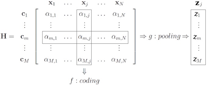
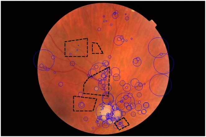
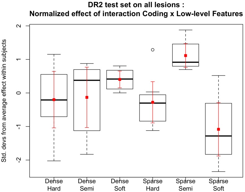
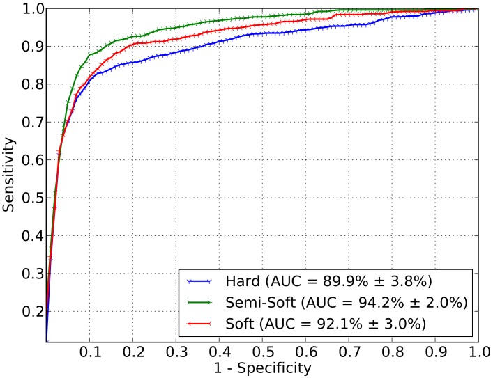
Diabetic Retinopathy (DR) is a complication of diabetes that can lead to blindness if not readily discovered. Automated screening algorithms have the potential to improve identification of patients who need further medical attention. However, the identification of lesions must be accurate to be useful for clinical application. The bag-of-visual-words (BoVW) algorithm employs a maximum-margin classifier in a flexible framework that is able to detect the most common DR-related lesions such as microaneurysms, cotton-wool spots and hard exudates. BoVW allows to bypass the need for pre- and post-processing of the retinographic images, as well as the need of specific ad hoc techniques for identification of each type of lesion. An extensive evaluation of the BoVW model, using three large retinograph datasets (DR1, DR2 and Messidor) with different resolution and collected by different healthcare personnel, was performed. The results demonstrate that the BoVW classification approach can identify different lesions within an image without having to utilize different algorithms for each lesion reducing processing time and providing a more flexible diagnostic system. Our BoVW scheme is based on sparse low-level feature detection with a Speeded-Up Robust Features (SURF) local descriptor, and mid-level features based on semi-soft coding with max pooling. The best BoVW representation for retinal image classification was an area under the receiver operating characteristic curve (AUC-ROC) of 97.8% (exudates) and 93.5% (red lesions), applying a cross-dataset validation protocol. To assess the accuracy for detecting cases that require referral within one year, the sparse extraction technique associated with semi-soft coding and max pooling obtained an AUC of 94.2 ± 2.0%, outperforming current methods. Those results indicate that, for retinal image classification tasks in clinical practice, BoVW is equal and, in some instances, surpasses results obtained using dense detection (widely believed to be the best choice in many vision problems) for the low-level descriptors.
Conflict of interest statement
Figures
References
-
- Sinthanayothin C, Boyce JF, Williamson TH, Cook HL, Mensah E, et al. (2002) Automated detection of diabetic retinopathy on digital fundus images. Diabetic Medicine: a Journal of the British Diabetic Association 19: 105–112. - PubMed
-
- Jelinek HF, Cree MJ, Worsley D, Luckie AP, Nixon P (2006) An automated microaneurysm detector as a tool for identification of diabetic retinopathy in rural optometric practice. Clinical and Experimental Optometry 89: 299–305. - PubMed
-
- Fleming AD, Philip S, Goatman KA, Olson JA, Sharp PF (2006) Automated microaneurysm detection using local contrast normalization and local vessel detection. IEEE Transactions on Medical Imaging 25: 1223–1232. - PubMed
-
- Niemeijer M, van Ginneken B, Russell SR, Suttorp-Schulten MSA, Abràmoff MD (2007) Automated detection and differentiation of drusen, exudates, and cotton-wool spots in digital color fundus photographs for diabetic retinopathy diagnosis. Investigative Ophthalmology & Visual Science 48: 2260–2267. - PMC - PubMed
-
- Giancardo L, Mériaudeau F, Karnowski TP, Tobin KW, Li Y, et al... (2010) Microaneurysms Detection with the Radon Cliff Operator in Retinal Fundus Images. In SPIE Medical Imaging. 76230U-76230U. International Society for Optics and Photonics.
Publication types
MeSH terms
LinkOut - more resources
Full Text Sources
Other Literature Sources
Medical
