Review
. 2014 May 30;15(6):9670-717.
doi: 10.3390/ijms15069670.

Computational and experimental approaches to reveal the effects of single nucleotide polymorphisms with respect to disease diagnostics

Affiliations
Review

Computational and experimental approaches to reveal the effects of single nucleotide polymorphisms with respect to disease diagnostics

Tugba G Kucukkal et al. Int J Mol Sci. .

Abstract

DNA mutations are the cause of many human diseases and they are the reason for natural differences among individuals by affecting the structure, function, interactions, and other properties of DNA and expressed proteins. The ability to predict whether a given mutation is disease-causing or harmless is of great importance for the early detection of patients with a high risk of developing a particular disease and would pave the way for personalized medicine and diagnostics. Here we review existing methods and techniques to study and predict the effects of DNA mutations from three different perspectives: in silico, in vitro and in vivo. It is emphasized that the problem is complicated and successful detection of a pathogenic mutation frequently requires a combination of several methods and a knowledge of the biological phenomena associated with the corresponding macromolecules.


Figures

Figure 1
Figure 1
Flowchart illustrating the methods (left) used for assessment of potential impacts (right) of DNA mutations on protein properties and interactions. nsSNP, non-synonymous Single Nucleotide Polymorphism.
Figure 2
Figure 2
Schematic diagram of different states involved in the energy calculations and the corresponding equations used to predict the change of the folding free energy upon mutation. WT, Wild-type; MT, Mutant; Green: Folded WT protein in vacuum (V, gray); Light Green: Folded WT protein in solvent (S, blue); Violet: Folded MT protein in vacuum; Light Violet: Folded MT protein in solvent, Unfolded (U) states are represented by black curved lines.
Figure 3
Figure 3
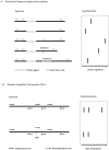
Principles of Restriction Fragment Length Polymorphisms (A) and Random amplified polymorphic DNA (B). The resulting fragments from target genome DNA are generated (left) and separated by gel electrophoresis followed by appropriate detection approach (right). To perform restriction fragment length polymorphisms (RFLP), genome DNA from variant individuals are subject to restriction digest (restriction sites are indicated by triangle) and random genomic probe (complement sequences are indicated by empty rectangle) is used for Southern Blot detection. Bands shown in Southern Blot represent (1) wild type; (2) loss of restriction site; (3) gain restriction site; (4) deletion of DNA fragment; (5) insertion of DNA fragment, from top to bottom. To perform RAPD, random primer sets are used to amplify genome DNA. Each matched primer set (full line arrow) generates a specific size product that appears in both wild type (top) and mutant (bottom) genome. Meanwhile mismatched primer (dashed line arrow) causes band absent in mutant.
Figure 4
Figure 4
(A) Percentage distribution of different Rett syndrome mutants. Three major types of sequence changes in specific domain are shown in the pie chart. Missense mutants in the Methyl-CpG-binding domain (MBD) and nonsense mutations that interrupt the transcription repression domain (TRD) are the major types. Synonymous mutations in exons and all mutants in the 5' UTR, the 3' UTR and introns are termed Silent; (B) The location and frequency of MECP2 Rett syndrome mutants. The most frequent 10 mutants are labeled. R106W, arginine to tryptophan point mutation at residue 106; R133C, arginine to cysteine point mutation at residue 133; T158M, threonine to methionine point mutation at residue 158; R168X, arginine to stop codon at residue 168; R255X, arginine to stop codon at residue 255; R294X, arginine to stop codon at residue 294; R306C, arginine to cysteine point mutation at residue 306; R9fs, frame-shift from arginine at residue 9; frame-shift from lysine at residue 386. The first eight mutants are termed mutational “hotspots”. Data from the IRSF MECP2 Gene Variation Database.

Similar articles

Cited by

References

    1. Potapov V., Cohen M., Schreiber G. Assessing computational methods for predicting protein stability upon mutation: good on average but not in the details. Protein Eng. Des. Sel. 2009;22:553–560. doi: 10.1093/protein/gzp030. - DOI - PubMed
    1. Thusberg J., Vihinen M. Pathogenic or Not? And if so, then how? Studying the effects of missense mutations using bioinformatics methods. Hum. Mutat. 2009;30:703–714. doi: 10.1002/humu.20938. - DOI - PubMed
    1. Gonzalez-Castejon M., Marin F., Soler-Rivas C., Reglero G., Visioli F., Rodriguez-Casado A. Functional non-synonymous polymorphisms prediction methods: Current approaches and future developments. Curr. Med. Chem. 2011;18:5095–5103. doi: 10.2174/092986711797636081. - DOI - PubMed
    1. Thiltgen G., Goldstein R.A. Assessing predictors of changes in protein stability upon mutation using self-consistency. PLoS One. 2012;7:e46084. doi: 10.1371/journal.pone.0046084. - DOI - PMC - PubMed
    1. Zhang Z., Miteva M.A., Wang L., Alexov E. Analyzing effects of naturally occurring missense mutations. Comput. Math. Methods Med. 2012;2012:805827:1–805827:15. - PMC - PubMed

Publication types

LinkOut - more resources