. 2014 Aug 15;392(2):324-33.
doi: 10.1016/j.ydbio.2014.05.014. Epub 2014 Jun 2.

Differential regulation of Gli proteins by Sufu in the lung affects PDGF signaling and myofibroblast development

Affiliations

Differential regulation of Gli proteins by Sufu in the lung affects PDGF signaling and myofibroblast development

Chuwen Lin et al. Dev Biol. .

Abstract

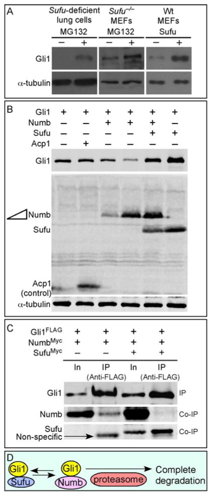
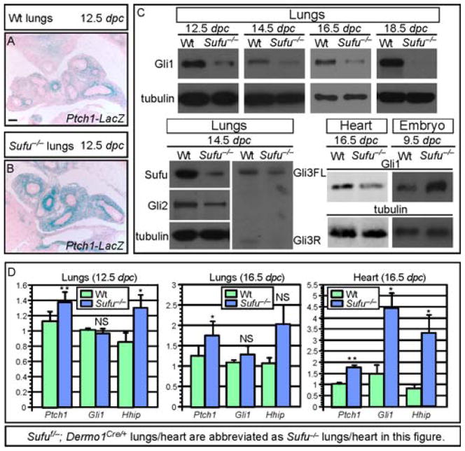
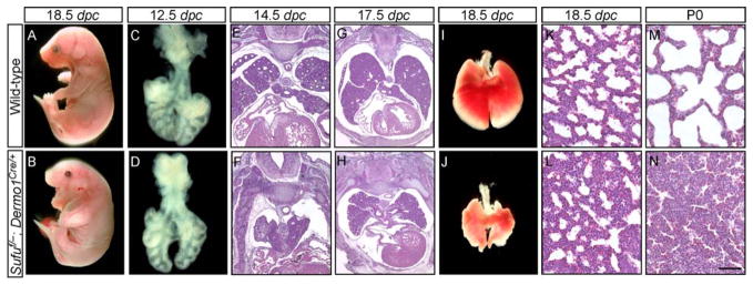
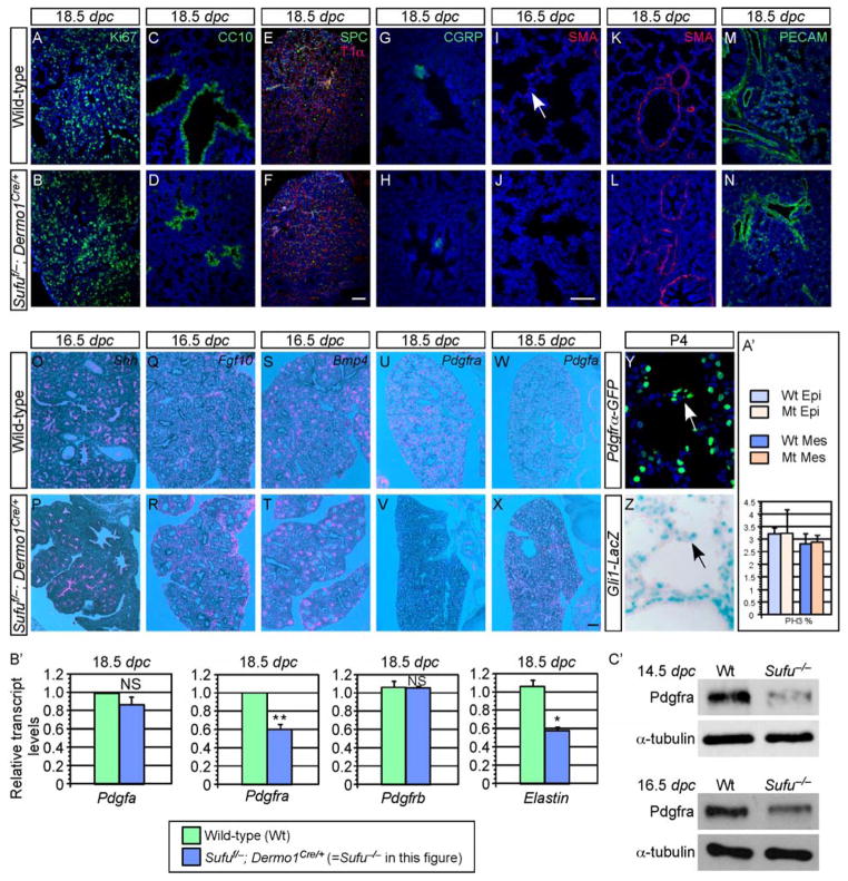
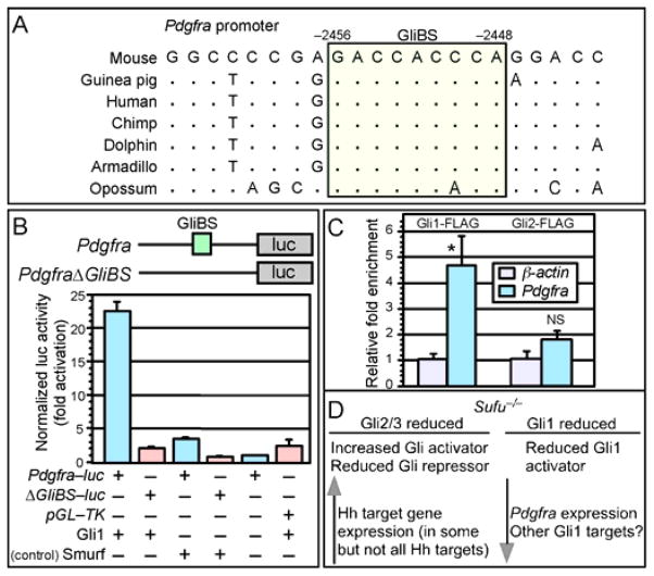
Mammalian Hedgehog (Hh) signaling relies on three Gli transcription factors to mediate Hh responses. This process is controlled in part by a major negative regulator, Sufu, through its effects on Gli protein level, distribution and activity. In this report, we showed that Sufu regulates Gli1 protein levels by antagonizing Numb/Itch. Otherwise, Numb/Itch would induce Gli1 protein degradation. This is in contrast to inhibition of Spop-mediated degradation of Gli2/3 by Sufu. Thus, controlling protein levels of all three Gli genes by Sufu is a conserved mechanism to modulate Hh responses albeit via distinct pathways. These findings in cell-based assays were further validated in vivo. In analyzing how Sufu controls Gli proteins in different tissues, we discovered that loss of Sufu in the lung exerts different effects on Hh target genes. Hh targets Ptch1/Hhip are upregulated in Sufu-deficient lungs, consistent with Hh pathway activation. Surprisingly, protein levels of Hh target Gli1 are reduced. We also found that myofibroblasts are absent from many prospective alveoli of Sufu-deficient lungs. Myofibroblast development is dependent on PDGF signaling. Interestingly, analysis of the Pdgfra promoter revealed a canonical Gli-binding site where Gli1 resides. These studies support a model in which loss of Sufu contributes to compromised Pdgfra activation and disrupts myofibroblast development in the lung. Our work illustrates the unappreciated complexity of Hh responses where distinct Hh targets could respond differently depending on the availability of Gli proteins that control their expression.

Keywords: Gli; Hedgehog; Lung; Myofibroblast; PDGF; Sufu.


Figures

Figure 1
Figure 1. Control of Gli1 protein levels by Sufu
(A) Western blot analysis of lysates from Sufu-deficient (Sufuf/−; Dermo1Cre/+) lung cells or Sufu null (Sufu−/−) MEFs treated with MG132 to block proteasome-mediated degradation. Endogenous Gli1 protein levels were elevated when protein degradation was inhibited in Sufu mutants. Similarly, protein levels of transfected Gli1 were increased when Sufu was co-expressed in wild-type (wt) MEFs. These results suggest that Sufu stabilizes Gli1 by preventing proteasome-dependent Gli1 degradation. Note that cycloheximide was added to block new protein synthesis in these studies. (B) Western blot analysis of lysates from HEK293T cells expressing various combinations of Gli1, Numb, Sufu and Acp1 (control). Numb expression resulted in reduction in Gli1 protein levels. This is consistent with previous reports in which Numb was shown to activate the E3 ligase Itch, leading to Gli1 ubiquitination and degradation. Numb-induced Gli1 reduction was reversed when Sufu was co-expressed with Numb. Tubulin was used as the loading control. (C) Western blot analysis of immunoprecipitated Gli1FLAG from HEK293T cell lysates to test the competition between Sufu and Numb in binding to Gli1. Co-immunoprecipitated NumbMyc by Gli1 was significantly reduced when SufuMyc was also pulled down by Gli1. (D) A model in which Sufu stabilizes Gli1 by inhibiting Numb-mediated protein degradation. In, input; IP, immunoprecipitation.
Figure 2
Figure 2. Upregulation of Hh target gene expression with concomitant Gli1 protein reduction in the absence of Sufu
(A–B) β-galactosidase staining of wild-type (wt) and Sufu-deficient (Sufuf/−; Dermo1Cre/+; Ptch1LacZ/+) lungs to detect Ptch1 expression in lung mesenchyme. Ptch1-LacZ expression was stronger and broader in Sufu mutants compared to wt. (C) Western blot analysis of endogenous Gli1 protein levels in wt and Sufu mutant lungs. Gli1 protein levels were decreased in Sufu mutant lungs collected at various stages of lung development. This resembles reduced protein levels of Gli2/3 in Sufu mutant lungs. Likewise, a reduction in Gli1 protein levels was also detected in Sufu-deficient hearts. (D) qPCR analysis of Ptch1 and Gli1 mRNA in wt and Sufu mutant lungs. Ptch1 and Hhip mRNA levels were elevated in Sufu mutant lungs while Gli1 mRNA levels were unaltered. This suggests that reduced Gli1 protein levels in Sufu-deficient lungs are a result of loss of Sufu and not due to changes in Gli1 transcript levels. All values are means ± standard deviation. * P < 0.05; ** P < 0.01; NS, not significant (unpaired Student’s t-test) (n=6 for 12.5 dpc lungs; n=4 for 16.5 dpc lungs; n=4 for 16.5 dpc heart). Elevation of Hhip mRNA levels at 16.5 dpc was not statistically significant likely due to large variations in transcript levels among different samples. Note that Sufuf/−; Dermo1Cre/+ lungs/heart are abbreviated as Sufu−/− lungs/heart while Sufu−/− embryos represent Sufu null embryos in this figure. FL, full-length; R, repressor. Scale bar = 100 μm for A–B.
Figure 3
Figure 3. Conditional inactivation of Sufu in lung mesenchyme
(A–N) External morphology (A, B), dissected lungs (C, D, I, J) and histology (E–H, K–N) of lung sections from wild-type (wt) and Sufuf/−; Dermo1Cre/+ mouse embryos at various embryonic stages and postnatal (p) day 0 as indicated. More than 50 Sufu mutants were examined. The phenotypes in Sufu mutant lungs were completely penetrant and showed little variation from animal to animal. Epithelial and mesenchymal development was defective in Sufu mutants, resulting in a smaller sized lung with a compact mesenchyme. The length of the proximal-distal axis of Sufu mutant lungs is ~80% of that of wt lungs. Sufu mutant mice cannot expand their lungs and died a few hours after birth. Early patterns of epithelial branching appeared to be established properly in Sufu-deficient lungs, leading to the correct number and positioning of lung lobes. Epithelial branching at later stages of development was not as extensive in Sufu-deficient lungs compared to that in wt lungs. dpc, days post coitus. Scale bars: K–N, 50 μm.
Figure 4
Figure 4. Disruption of myofibroblast development in Sufu-deficient lungs
(A–Z) Immunostaining (A–N, Y, Z) and in situ hybridization (O–X) of wild-type (wt) and Sufu-deficient (Sufuf/−; Dermo1Cre/+) lungs. No apparent difference in cell proliferation rate (judged by Ki67 and PH3 staining) or cell death was found between wt (A) and Sufu mutant (B) lungs. Quantification of cell proliferation in the epithelial and mesenchymal compartments at 12.5 dpc was shown in A′. Major epithelial cell types, including Clara cells (CC10+) (C, D), alveolar type II (SPC+), type I (T1α+) cells (E, F) and pulmonary neuroendocrine cells (CGRP+) (G, H) were properly specified in the absence of Sufu. Most mesenchymal cell types such as the bronchial smooth muscle (SMA+) (K, L) and blood vessels (PECAM+) (M, N) were also properly generated. By contrast, myofibroblasts (white arrow; marked by smooth muscle actin [SMA] staining) were greatly reduced in Sufu mutant lungs (I). This was associated with decreased Pdgfra expression (pink signal) (compare V to U) while Pdgf ligand expression was unaltered (compare X to W). In addition, the expression patterns and levels of Shh (O, P), Fgf10 (Q, R) and Bmp4 (S, T) or other components in Hh, Fgf and Bmp signaling were similar between wt and Sufu mutant lungs. Both Gli1 and Pdgfra were detected in the secondary septa of alveoli (arrows in Y, Z). (B′) qPCR analysis of Pdgfa, Pdgfra, Pdgfrb and Elastin mRNA in wt and Sufu mutant lungs. Pdgfra mRNA levels were reduced in Sufu mutant lungs while Pdgfa and Pdgfrb mRNA levels were unaltered. This is consistent with results from in situ hybridization. Elastin transcript levels were also reduced in Sufu mutant lungs, consistent with defective myofibroblast development. All values are means ± standard deviation. * P < 0.05; ** P < 0.01; NS, not significant (unpaired Student’s t-test) (n=3). (C′) Western blot of endogenous Pdgfra protein levels in wt and Sufu mutant lungs. Pdgfra protein levels were decreased in Sufu mutant lungs collected at various stages of lung development. This was likely due to reduced Pdgfra transcript levels in Sufu mutant lungs. dpc, days post coitus. Scale bars: A–F and K–N, 50 μm; G–J and Y–Z, 50 μm; O–X, 100 μm.
Figure 5
Figure 5. Gli1 and regulation of Pdgfra promoter activity
(A) Sequence analysis of the Pdgfra promoter from different species. A conserved canonical Gli-binding site (GliBS) is boxed and colored. The numbers represent distances from the transcriptional start site of Pdgfra, which is marked as position zero. (B) Schematic diagram depicting Pdgfra-luc reporter constructs in which mouse Pdgfra promoter fragments are placed upstream of firefly luciferase (luc). A canonical GliBS is present in the Pdgfra promoter and is mutated in the control construct PdgfraΔGliBS-luc (abbreviated as ΔGliBS-luc in the figure). Addition of Gli1 activated Pdgfra-luc and not PdgfraΔGliBS-luc in cell-based assays. Control nuclear protein Smurf did not induce Pdgfra-luc expression. (C) Gli1 but not Gli2 occupied the Pdgfra promoter by ChIP analysis using MEFs expressing FLAG-tagged Gli1 and Gli2. Gli-binding on the Pdgfra promoter was normalized to the β-actin control promoter. All values are means ± standard deviation. * P < 0.05; NS, not significant (unpaired Student’s t-test) (n=3). (D) A model of differential regulation of Gli proteins by Sufu. Loss of Sufu results in reduced protein levels of all three Gli proteins. Reduction in Gli2/3 protein levels is associated with increased Gli activators and reduced Gli repressors. This would lead to overall Hh pathway activation and Hh target gene expression. By contrast, reduced protein levels of the constitutive activator Gli1 result in downregulation of Hh targets that primarily rely on Gli1 for their expression such as Pdgfra in the lung.

Similar articles

Cited by

References

    1. Ausubel FM, Brent R, Kingston RE, Moore DD, Seidman JG, Smith JA, Struhl K. Current Protocols in Molecular Biology (Wiley) 2003
    1. Bai CB, Auerbach W, Lee JS, Stephen D, Joyner AL. Gli2, but not Gli1, is required for initial Shh signaling and ectopic activation of the Shh pathway. Development. 2002;129:4753–4761. - PubMed
    1. Bai CB, Stephen D, Joyner AL. All mouse ventral spinal cord patterning by hedgehog is Gli dependent and involves an activator function of Gli3. Dev Cell. 2004;6:103–115. - PubMed
    1. Barakat MT, Humke EW, Scott MP. Learning from Jekyll to control Hyde: Hedgehog signaling in development and cancer. Trends Mol Med. 2010;16:337–348. - PMC - PubMed
    1. Barnfield PC, Zhang X, Thanabalasingham V, Yoshida M, Hui CC. Negative regulation of Gli1 and Gli2 activator function by Suppressor of fused through multiple mechanisms. Differentiation; research in biological diversity. 2005;73:397–405. - PubMed

Publication types

MeSH terms