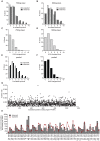
High throughput mutagenesis for identification of residues regulating human prostacyclin (hIP) receptor expression and function
- PMID: 24886841
- PMCID: PMC4041722
- DOI: 10.1371/journal.pone.0097973
High throughput mutagenesis for identification of residues regulating human prostacyclin (hIP) receptor expression and function
Abstract
The human prostacyclin receptor (hIP receptor) is a seven-transmembrane G protein-coupled receptor (GPCR) that plays a critical role in vascular smooth muscle relaxation and platelet aggregation. hIP receptor dysfunction has been implicated in numerous cardiovascular abnormalities, including myocardial infarction, hypertension, thrombosis and atherosclerosis. Genomic sequencing has discovered several genetic variations in the PTGIR gene coding for hIP receptor, however, its structure-function relationship has not been sufficiently explored. Here we set out to investigate the applicability of high throughput random mutagenesis to study the structure-function relationship of hIP receptor. While chemical mutagenesis was not suitable to generate a mutagenesis library with sufficient coverage, our data demonstrate error-prone PCR (epPCR) mediated mutagenesis as a valuable method for the unbiased screening of residues regulating hIP receptor function and expression. Here we describe the generation and functional characterization of an epPCR derived mutagenesis library compromising >4000 mutants of the hIP receptor. We introduce next generation sequencing as a useful tool to validate the quality of mutagenesis libraries by providing information about the coverage, mutation rate and mutational bias. We identified 18 mutants of the hIP receptor that were expressed at the cell surface, but demonstrated impaired receptor function. A total of 38 non-synonymous mutations were identified within the coding region of the hIP receptor, mapping to 36 distinct residues, including several mutations previously reported to affect the signaling of the hIP receptor. Thus, our data demonstrates epPCR mediated random mutagenesis as a valuable and practical method to study the structure-function relationship of GPCRs.
Conflict of interest statement
Figures


Similar articles
-
Arginine (CGC) codon targeting in the human prostacyclin receptor gene (PTGIR) and G-protein coupled receptors (GPCR).Gene. 2007 Jul 1;396(1):180-7. doi: 10.1016/j.gene.2007.03.016. Epub 2007 Apr 1. Gene. 2007. PMID: 17481829 Free PMC article.
-
New insights into human prostacyclin receptor structure and function through natural and synthetic mutations of transmembrane charged residues.Br J Pharmacol. 2007 Oct;152(4):513-22. doi: 10.1038/sj.bjp.0707413. Epub 2007 Aug 20. Br J Pharmacol. 2007. PMID: 17704830 Free PMC article.
-
Lessons from diversity of directed evolution experiments by an analysis of 3,000 mutations.Biotechnol Bioeng. 2014 Dec;111(12):2380-9. doi: 10.1002/bit.25302. Epub 2014 Jul 14. Biotechnol Bioeng. 2014. PMID: 24904008
-
Human prostacyclin receptor structure and function from naturally-occurring and synthetic mutations.Prostaglandins Other Lipid Mediat. 2007 Jan;82(1-4):95-108. doi: 10.1016/j.prostaglandins.2006.05.010. Epub 2006 Jul 3. Prostaglandins Other Lipid Mediat. 2007. PMID: 17164137 Review.
-
Polishing the craft of genetic diversity creation in directed evolution.Biotechnol Adv. 2013 Dec;31(8):1707-21. doi: 10.1016/j.biotechadv.2013.08.021. Epub 2013 Sep 6. Biotechnol Adv. 2013. PMID: 24012599 Review.
Cited by
-
High Throughput Random Mutagenesis and Single Molecule Real Time Sequencing of the Muscle Nicotinic Acetylcholine Receptor.PLoS One. 2016 Sep 20;11(9):e0163129. doi: 10.1371/journal.pone.0163129. eCollection 2016. PLoS One. 2016. PMID: 27649498 Free PMC article.
-
Variomics screen identifies the re-entrant loop of the calcium-activated chloride channel ANO1 that facilitates channel activation.J Biol Chem. 2015 Jan 9;290(2):889-903. doi: 10.1074/jbc.M114.618140. Epub 2014 Nov 25. J Biol Chem. 2015. PMID: 25425649 Free PMC article.
-
OverFlap PCR: A reliable approach for generating plasmid DNA libraries containing random sequences without a template bias.PLoS One. 2022 Aug 8;17(8):e0262968. doi: 10.1371/journal.pone.0262968. eCollection 2022. PLoS One. 2022. PMID: 35939421 Free PMC article.
-
GigaAssay - An adaptable high-throughput saturation mutagenesis assay platform.Genomics. 2022 Jul;114(4):110439. doi: 10.1016/j.ygeno.2022.110439. Epub 2022 Jul 26. Genomics. 2022. PMID: 35905834 Free PMC article.
-
High-throughput mutagenesis using a two-fragment PCR approach.Sci Rep. 2017 Jul 28;7(1):6787. doi: 10.1038/s41598-017-07010-4. Sci Rep. 2017. PMID: 28754896 Free PMC article.
References
-
- Narumiya S, Sugimoto Y, Ushikubi F (1999) Prostanoid receptors: structures, properties, and functions. Physiol Rev 79: 1193–1226. - PubMed
-
- Giguere V, Gallant MA, de Brum-Fernandes AJ, Parent JL (2004) Role of extracellular cysteine residues in dimerization/oligomerization of the human prostacyclin receptor. Eur J Pharmacol 494: 11–22. - PubMed
-
- Stitham J, Gleim SR, Douville K, Arehart E, Hwa J (2006) Versatility and differential roles of cysteine residues in human prostacyclin receptor structure and function. J Biol Chem 281: 37227–37236. - PubMed
-
- Egan KM, Lawson JA, Fries S, Koller B, Rader DJ, et al. (2004) COX-2-derived prostacyclin confers atheroprotection on female mice. Science 306: 1954–1957. - PubMed
-
- Xiao CY, Hara A, Yuhki K, Fujino T, Ma H, et al. (2001) Roles of prostaglandin I(2) and thromboxane A(2) in cardiac ischemia-reperfusion injury: a study using mice lacking their respective receptors. Circulation 104: 2210–2215. - PubMed
Publication types
MeSH terms
Substances
LinkOut - more resources
Full Text Sources
Other Literature Sources

