. 2014 Jun 2;9(6):e98239.
doi: 10.1371/journal.pone.0098239. eCollection 2014.

Functional effects of Toll-like receptor (TLR)3, 7, 9, RIG-I and MDA-5 stimulation in nasal epithelial cells

Affiliations

Functional effects of Toll-like receptor (TLR)3, 7, 9, RIG-I and MDA-5 stimulation in nasal epithelial cells

Lotta Tengroth et al. PLoS One. .

Abstract

Background: The human nasal epithelium is an important physical barrier, and a part of the innate immune defense that protect against pathogens. The epithelial cells recognize microbial components by pattern-recognition receptors (PRRs), and thereby trigger an immune response. Even though TLR3, TLR7, TLR9, RIG-I and MDA-5 are all known to respond to viral stimulation, their potential role in chronic airway inflammation triggered by local cytokine release remains to be established.

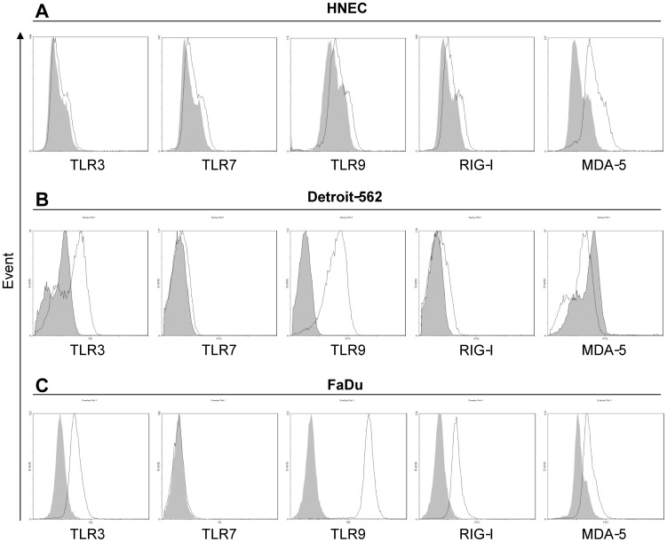
Methods: mRNA and corresponding protein expression of TLR3, TLR7, TLR9, RIG-I and MDA-5 were analyzed in nasal biopsies and various upper airway epithelial cell lines using real-time reverse transcription PCR, immunohistochemistry and flow cytometry. Ligand induced, cytokine release, was evaluated with ELISA.

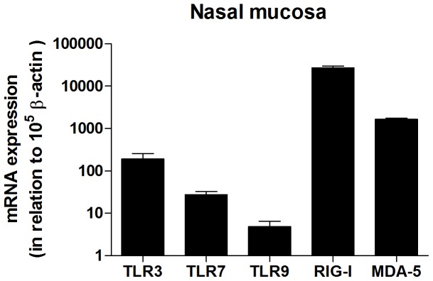
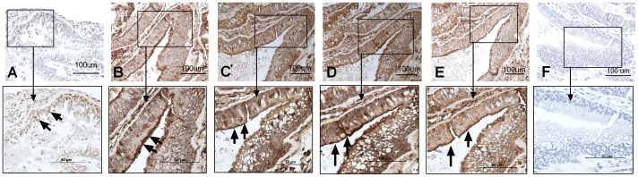
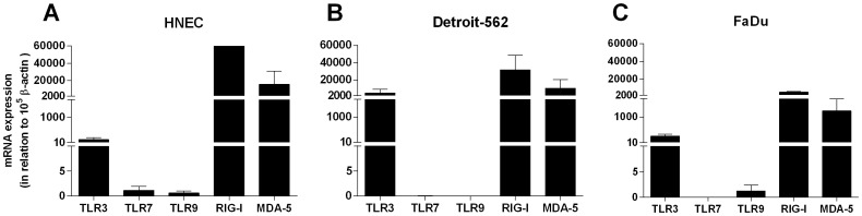
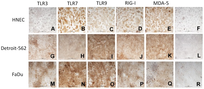
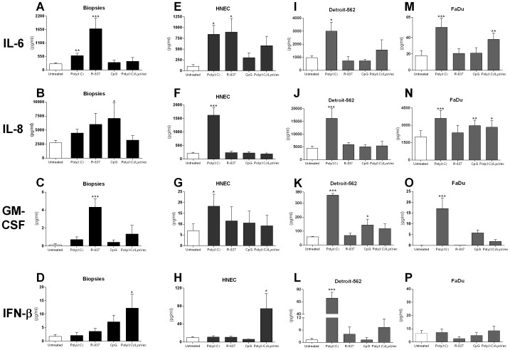
Results: Nasal biopsies were found to express TLR3, TLR7, TLR9, RIG-I and MDA-5, with the most abundant expression in the surface epithelium. These receptors were verified in primary human nasal epithelial cell (HNEC) as well as in the airway epithelial cell lines Detroit-562 and FaDu. Poly(I:C) (TLR3) and R-837 (TLR7) stimulation increased secretion of IL-6 and GM-CSF from the nasal mucosa and the epithelial cell lines. CpG (TLR9) stimulation caused release of IL-8 in the nasal mucosa and in FaDu. Poly(I:C)/LyoVec (RIG-I/MDA-5) stimulation activated the secretion of IFN-β in the nasal mucosa. A corresponding release was also detected from HNEC and Detroit-562.

Conclusion: The nasal epithelium has the ability to recognize viral intrusion through TLR and RLR receptors, and the subsequent response might have a role in exacerbation of inflammatory diseases like allergic rhinitis and chronic rhinosinusitis.


Conflict of interest statement

Competing Interests: The authors have declared that no competing interests exist.

Figures

Figure 1
Figure 1. mRNA expression of TLR3, TLR7, TLR9, RIG-I and MDA-5 in human nasal mucosa.
mRNA expression in nasal mucosal biopsies was determined by real-time reverse transcriptase-PCR (n = 20). Data is presented in relation to β-actin as 2-ΔCt × 105 and depicted in log scale as mean ± SEM.
Figure 2
Figure 2. The nasal epithelium expresses TLR3, TLR7, TLR9, RIG-I and MDA-5.
Sections of nasal biopsies were incubated with antibodies against TLR3 (A), TLR7 (B), TLR9 (C), RIG-I (D), and MDA-5 (E) and visualized by 3, 3′-diaminobenzidine (brown). In control slides (F), N-series universal negative control reagent was used. All sections were accompanied with a square magnification. All slides were counterstained with haematoxylin (blue). The figure shows one representative biopsy out of four (3 male, 1 female). The arrows indicate positive stained cells.
Figure 3
Figure 3. mRNA expression of TLR3, TLR7, TLR9, RIG-I and MDA-5 in epithelial cells.
Levels of innate immune receptors in human nasal epithelial cells (HNEC) (n = 5) (A), Detroit-562 (n = 6) (B), and FaDu (n = 4) (C) was determined by real-time reverse transcriptase-PCR. Data is presented in relation to β-actin as 2-ΔCt × 105 and depicted in linear scale as mean ± SEM.
Figure 4
Figure 4. Expression of TLR3, TLR7, TLR9, RIG-I and MDA-5 on epithelial cells.
Epithelial cells from primary HNEC (A–E), Detroit-562 (G–K) and FaDu (M–Q) were incubated with antibodies against TLR3, TLR7, TLR9, RIG-I, and MDA-5 and visualized by 3, 3′-diaminobenzidine (brown). In controls, N-series universal negative control reagent was used (F, L, R). All cells were counterstained with haematoxylin (blue). The figure shows one representative staining out of three independent experiments. The markers in the figure are 50 µm.
Figure 5
Figure 5. Expression of TLR3, TLR7, TLR9, RIG-I and MDA-5 proteins on epithelial cells.
HNEC (A), Detroit-562 (B) and FaDu (C) were stained intracellularly with Abs against TLR3, TLR7, TLR9, RIG-I and MDA-5 (open histograms) or appropriate isotype control (shaded histograms) and analyzed by flow cytometry. Representative pictures from one out of three independent experiments are shown.
Figure 6
Figure 6. TLR3, TLR7, TLR9, RIG-I and MDA-5 stimulation promotes cytokine release.
Nasal biopsies and epithelial cells were cultured in the absence (Untreated) or presence of 10 µg/ml poly(I:C) (TLR3), 10 µg/ml R-837 (TLR7), 1 µM CpG (TLR9) and 1 µg/ml poly(I:C)/LyoVec (RIG-I/MDA-5). TNF-α (10 ng/ml) was used as a positive control (data not shown). After 24 h, supernatants from nasal biopsies (n = 5) (A–D), HNEC (n = 6–9) (E–H), Detroit-562 (n = 5) (I–L) and FaDu (n = 5–9) (M–P) were collected and analyzed for levels of IL-6, IL-8, GM-CSF and IFN-β using ELISA. Data is presented as mean ± SEM of 5 to 9 independent experiments. *, p<0.05; **, p<0.01; ***, p<0.001.

References

    1. Holgate ST (2007) Epithelium dysfunction in asthma. Journal of Allergy and Clinical Immunology 120: 1233–1246. - PubMed
    1. Schleimer RP, Kato A, Kern R, Kuperman D, Avila PC (2007) Epithelium: At the interface of innate and adaptive immune responses. Journal of Allergy and Clinical Immunology 120: 1279–1284. - PMC - PubMed
    1. Bals R, Hiemstra PS (2004) Innate immunity in the lung: how epithelial cells fight against respiratory pathogens. European Respiratory Journal 23: 327–333. - PubMed
    1. Kawai T, Akira S (2006) TLR signaling. Cell Death and Differentiation 13: 816–825. - PubMed
    1. Takeuchi O, Akira S (2010) Pattern recognition receptors and inflammation. Cell 140: 805–820. - PubMed

Publication types

MeSH terms

LinkOut - more resources