. 2014 Jun 2;9(6):e98365.
doi: 10.1371/journal.pone.0098365. eCollection 2014.

Structural and mutagenic analysis of the RM controller protein C.Esp1396I

Affiliations

Structural and mutagenic analysis of the RM controller protein C.Esp1396I

Richard N A Martin et al. PLoS One. .

Abstract

Bacterial restriction-modification (RM) systems are comprised of two complementary enzymatic activities that prevent the establishment of foreign DNA in a bacterial cell: DNA methylation and DNA restriction. These two activities are tightly regulated to prevent over-methylation or auto-restriction. Many Type II RM systems employ a controller (C) protein as a transcriptional regulator for the endonuclease gene (and in some cases, the methyltransferase gene also). All high-resolution structures of C-protein/DNA-protein complexes solved to date relate to C.Esp1396I, from which the interactions of specific amino acid residues with DNA bases and/or the phosphate backbone could be observed. Here we present both structural and DNA binding data for a series of mutations to the key DNA binding residues of C.Esp1396I. Our results indicate that mutations to the backbone binding residues (Y37, S52) had a lesser affect on DNA binding affinity than mutations to those residues that bind directly to the bases (T36, R46), and the contributions of each side chain to the binding energies are compared. High-resolution X-ray crystal structures of the mutant and native proteins showed that the fold of the proteins was unaffected by the mutations, but also revealed variation in the flexible loop conformations associated with DNA sequence recognition. Since the tyrosine residue Y37 contributes to DNA bending in the native complex, we have solved the structure of the Y37F mutant protein/DNA complex by X-ray crystallography to allow us to directly compare the structure of the DNA in the mutant and native complexes.


Conflict of interest statement

Competing Interests: The authors have declared that no competing interests exist.

Figures

Figure 1
Figure 1. Gene arrangement of the Esp1396I RM system and C.Esp1396I binding sites.
(A): The C-protein (C), endonuclease (R) and methyltransferase (M) genes. C/R genes form an operon that is convergent with the M gene. The three C-protein binding sites are shown in orange. (B) The OM operator controls expression of the M gene. (C) The OL and OR operators control the expression of the C/R genes.
Figure 2
Figure 2. Binding analysis and location of mutations.
(A) DNA binding curves of C.Esp1396I mutant proteins binding to the OM operator site from SPR data. (B) The amino acid residues mutated in this study (T36, Y37, R46 and S52) showing their location at the DNA-protein interface. Image generated in PyMOL using the wild type C.Esp1396I-19OM co-crystal structure (3UFD).
Figure 3
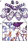
Figure 3. Coordination of the sulphate ion required in the crystal structure.
Top panel shows the asymmetric unit of the C.Esp1396I high resolution wild type X-ray crystal structure (4I6R) in orange with a symmetry related dimer in light orange. The sulphate ion is shown in pink. The zoom shows atoms hydrogen bonding directly to the sulphate including water molecules (dark blue spheres). Hydrogen bonds are shown as grey dashed lines.
Figure 4
Figure 4. Conformation of the flexible loop of C.Esp1396I.
(A): The three observed loop conformations observed in crystal structures of the native protein. The two alternative C.Esp1396I loop positions in 3G5G are shown in light blue (conformation I) and light green (conformation II) and the high resolution wild type loop position from 4I6R is shown in orange (III). (B): The side chains of residues S45, R46 and N47 are displayed as sticks.
Figure 5
Figure 5. Dimerisation contacts in the alternative loop conformations.
The high resolution wild type crystal structure (4I6R) is shown in orange (one monomer dark and one light) and the low resolution wild type is shown in blue/silver (one monomer dark and one light). The hydrogen bond is shown as a grey dashed line.
Figure 6
Figure 6. Conformation of the flexible loop of C.Esp1396I in the mutant crystal structures.
The three wild type loop conformations observed in 3G5G and 4I6R are shown in the same colours as in Figure 5 and are translucent. (A) T36A loop positions shown in green. (B) Y37A loop position shown in dark red. (C) Loop positions in both R46A structures shown in red (monoclinic space group) and purple (trigonal space group). (D) Loop position of S52A shown in light green. (E) Loop positions in Y37F (blue).
Figure 7
Figure 7. Comparison of the wild-type and Y37F mutant 19OM co-crystal structures.
The wild type structure is shown in pale blue and the Y37F mutant structure in purple. (A) An overlay of the two structures shows the high degree of similarity between the two. (B) The DNA “pinching” point at the central TATA sequence. Hydrogen bonds are shown as grey lines.
Figure 8
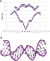
Figure 8. DNA bending analysis of the DNA from the C.Esp1396I-Y37F-19OM co-crystal structure.
(A) Groove width analysis of the DNA in the Y37F-19OM crystal structure. The wild-type 19OM co-crystal structure is shown in grey and the Y37F mutant co-crystal structure in purple. Major grooves widths are shown with squares and minor groove widths with circles. (B) The DNA duplex from the Y47F-19OM co-crystal structure shown with the helical axis in grey. Analysis performed using the Curves+ server .

References

    1. Wilson GG, Murray NE (1991) Restriction and modification systems. Annual Reviews in Genetics 25: 585–627. - PubMed
    1. Loenen WA, Dryden DT, Raleigh EA, Wilson GG, Murray NE (2014) Highlights of the DNA cutters: a short history of the restriction enzymes. Nucleic Acids Res. 42: 3–19. - PMC - PubMed
    1. Tao T, Bourne JC, Blumenthal RM (1991) A family of regulatory genes associated with type II restriction-modification systems. J. Bacteriol. 173: 1367–1375. - PMC - PubMed
    1. Ives CL, Nathan PD, Brooks JE (1992) Regulation of the BamHI restriction-modification system by a small intergenic open reading frame, bamHIC, in both Escherichia coli and Bacillus subtilis. J. Bacteriol. 174: 7194–7201. - PMC - PubMed
    1. Rimseliene R, Vaisvila R, Janulaitis A (1995) The eco72IC gene specifies a trans-acting factor which influences expression of both DNA methyltransferase and endonuclease from the Eco72I restriction-modification system. Gene 157: 217–219. - PubMed

Publication types

LinkOut - more resources