

Contribution of endothelial injury and inflammation in early phase to vein graft failure: the causal factors impact on the development of intimal hyperplasia in murine models
- PMID: 24887332
- PMCID: PMC4041877
- DOI: 10.1371/journal.pone.0098904
Contribution of endothelial injury and inflammation in early phase to vein graft failure: the causal factors impact on the development of intimal hyperplasia in murine models
Abstract
Objectives: Autologous veins are preferred conduits in by-pass surgery. However, long-term results are hampered by limited patency due to intimal hyperplasia. Although mechanisms involved in development of intimal hyperplasia have been established, the role of inflammatory processes is still unclear. Here, we studied leukocyte recruitment and intimal hyperplasia in inferior vena cava grafts transferred to abdominal aorta in mice.
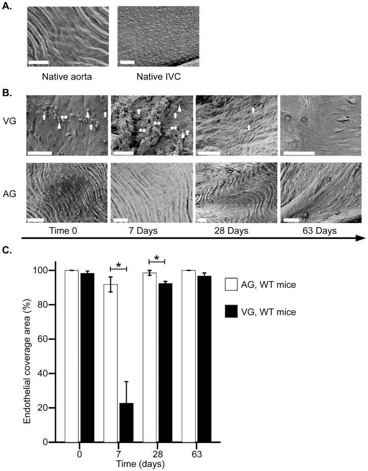
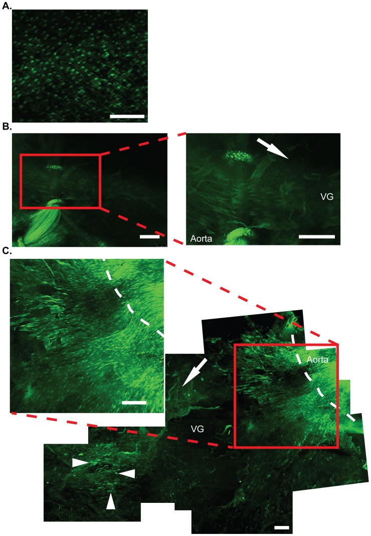
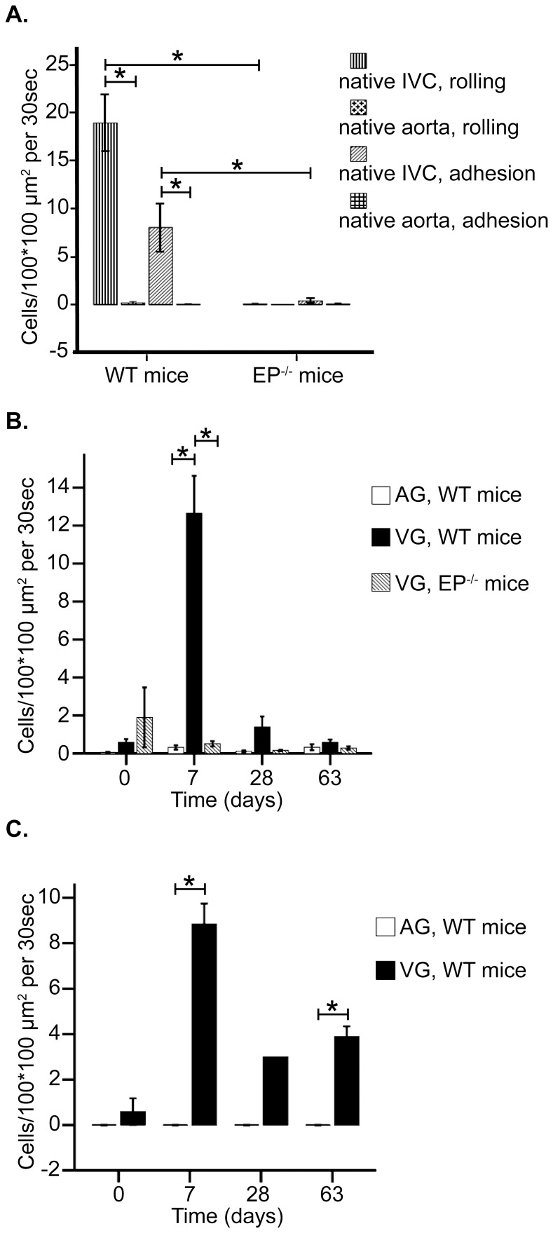
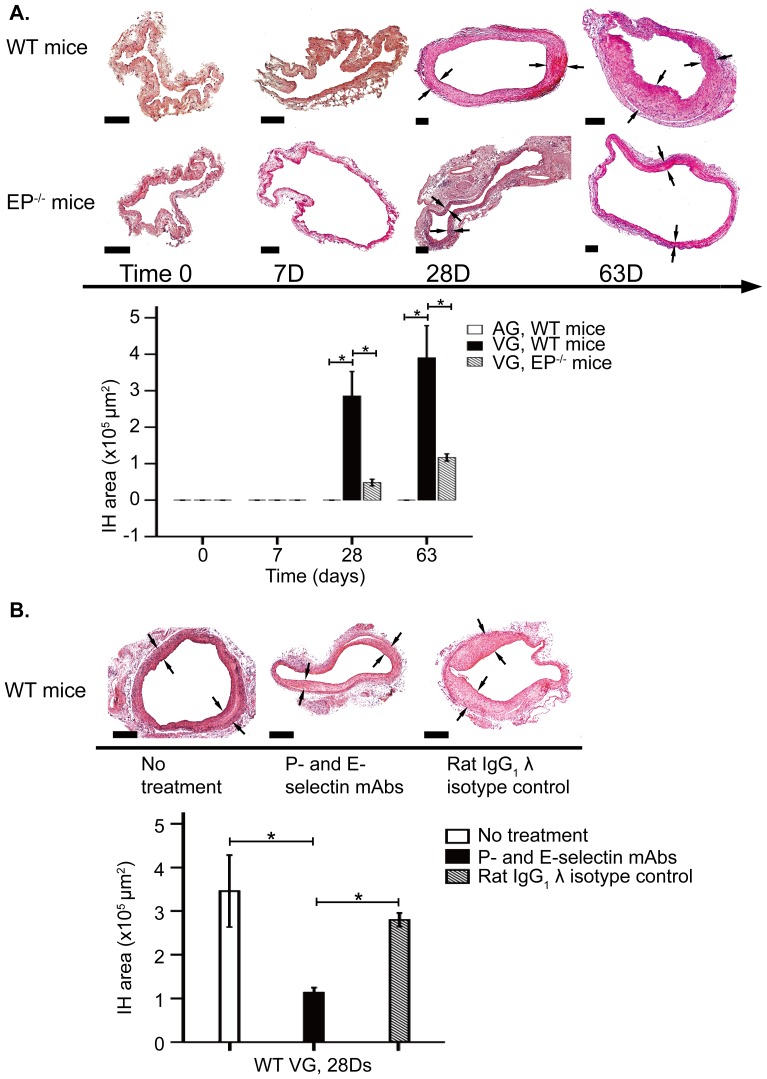
Methods and results: Several microscopic techniques were used to study endothelium denudation and regeneration and leukocyte recruitment on endothelium. Scanning electron microscopy demonstrated denudation of vein graft endothelium 7 days post-transfer and complete endothelial regeneration by 28 days. Examination of vein grafts transferred to mice transgenic for green fluorescent protein under Tie2 promoter in endothelial cells showed regeneration of graft endothelium from the adjacent aorta. Intravital microscopy revealed recruitment of leukocytes in vein grafts at 7 days in wild type mice, which had tapered off by 28 days. At 28 and 63 days there was significant development of intimal hyperplasia. In contrast; no injury, leukocyte recruitment nor intimal hyperplasia occurred in arterial grafts. Leukocyte recruitment was reduced in vein grafts in mice deficient in E- and P-selectin. In parallel, intimal hyperplasia was reduced in vein grafts in mice deficient in E- and P-selectin and in wild type mice receiving P-selectin/E-selectin function-blocking antibodies.
Conclusion: The results show that early phase endothelial injury and inflammation are crucial processes in intimal hyperplasia in murine vein grafts. The data implicate endothelial selectins as targets for intervention of vein graft disease.
Conflict of interest statement
Figures
References
-
- Cooley BC (1998) History of vein grafting. Microsurgery 18: 234–236. - PubMed
-
- Ehsan A, Mann MJ, Dell'Acqua G, Tamura K, Braun-Dullaeus R, et al. (2002) Endothelial healing in vein grafts: proliferative burst unimpaired by genetic therapy of neointimal disease. Circulation 105: 1686–1692. - PubMed
-
- Grondin CM (1984) Late results of coronary artery grafting: is there a flag on the field? J Thorac Cardiovasc Surg 87: 161–166. - PubMed
-
- Thatte HS, Khuri SF (2001) The coronary artery bypass conduit: I. Intraoperative endothelial injury and its implication on graft patency. Ann Thorac Surg 72: S2245–2252 discussion S2267–2270. - PubMed
-
- Deb S, Cohen EA, Singh SK, Une D, Laupacis A, et al. (2012) Radial artery and saphenous vein patency more than 5 years after coronary artery bypass surgery: results from RAPS (Radial Artery Patency Study). J Am Coll Cardiol 60: 28–35. - PubMed
Publication types
MeSH terms
Substances
LinkOut - more resources
Full Text Sources
Other Literature Sources
Miscellaneous
