. 2014 Jun 2;9(6):e98565.
doi: 10.1371/journal.pone.0098565. eCollection 2014.

Human umbilical cord-derived mesenchymal stem cells do not undergo malignant transformation during long-term culturing in serum-free medium

Affiliations

Human umbilical cord-derived mesenchymal stem cells do not undergo malignant transformation during long-term culturing in serum-free medium

Gecai Chen et al. PLoS One. .

Abstract

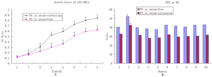
Background: Human umbilical cord-derived mesenchymal stem cells (hUC-MSCs) are in the foreground as a preferable application for treating diseases. However, the safety of hUC-MSCs after long-term culturing in vitro in serum-free medium remains unclear.

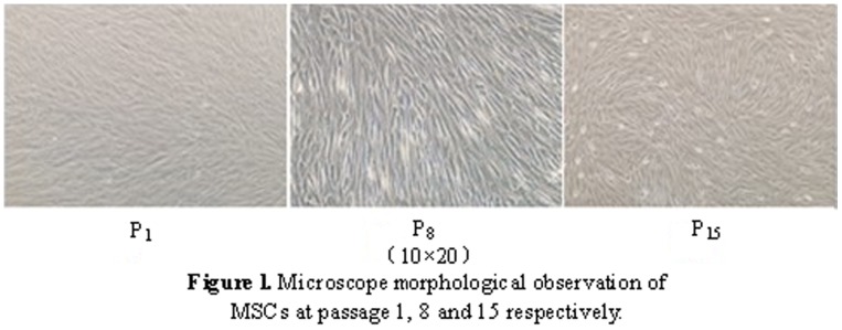
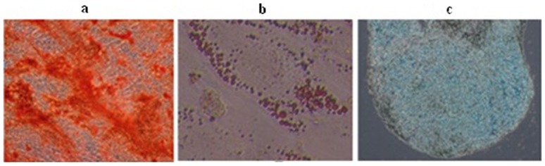
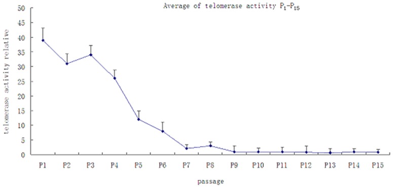
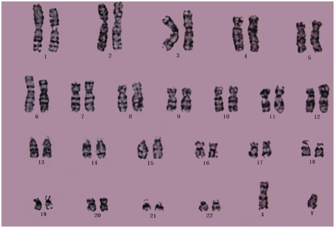
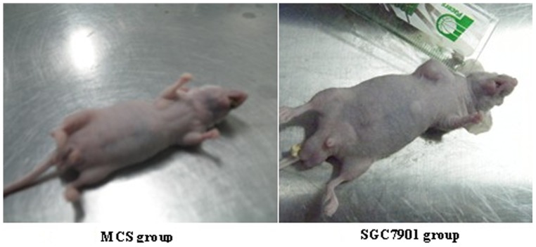
Methods: hUC-MSCs were separated by adherent tissue culture. hUC-MSCs were cultured in serum-free MesenCult-XF medium and FBS-bases DMEM complete medium. At the 1st, 3rd, 5th, 8th, 10th, and 15th passage, the differentiation of MSCs into osteogenic, chondrogenic, and adipogenic cells was detected, and MTT, surface antigens were measured. Tumorigenicity was analyzed at the 15th passage. Conventional karyotyping was performed at passage 0, 8, and 15. The telomerase activity of hUC-MSCs at passage 1-15 was analyzed.

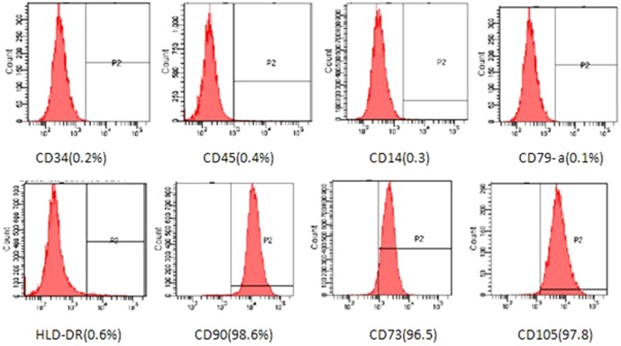
Results: Flow cytometry analysis showed that very high expression was detected for CD105, CD73, and CD90 and very low expression for CD45, CD34, CD14, CD79a, and HLA-DR. MSCs could differentiate into osteocytes, chondrocytes, and adipocytes in vitro. There was no obvious chromosome elimination, displacement, or chromosomal imbalance as determined from the guidelines of the International System for Human Cytogenetic Nomenclature. Telomerase activity was down-regulated significantly when the culture time was prolonged. Further, no tumors formed in rats injected with hUC-MSCs (P15) cultured in serum-free and in serum-containing conditions.

Conclusion: Our data showed that hUC-MSCs met the International Society for Cellular Therapy standards for conditions of long-term in vitro culturing at P15. Since hUC-MSCs can be safely expanded in vitro and are not susceptible to malignant transformation in serum-free medium, these cells are suitable for cell therapy.


Conflict of interest statement

Competing Interests: The authors have read the journal's policy and have the following conflicts: Co-author Aihuan Yue is employed by Jiangsu Beike Bio-Technology Co., Ltd. This does not alter the authors' adherence to PLOS ONE policies on sharing data and materials.

Figures

Figure 1
Figure 1. Microscope morphological observation of MSCs at passage 1, 8, and 15 from in serum-free condition.
Figure 2
Figure 2. Analysis of MSCs for the expression of surface markers by flow cytometric analysis.
The immunofluorescence analysis of MSCs was conducted at the 15th passage of cells obtained from donor 3.
Figure 3
Figure 3. Differentiation of hUC-MSCs from donor 3 was showed at the 15th passage.
(a) Alizarin Red S staining to detect osteogenesis differentiation. (b) Oil Red O staining to detect adipogenesis differentiation. (c) Alcian Blue staining to detect chondrogenic differentiation.
Figure 4
Figure 4. The proliferation capacity of hUC-MSCs
: (a).Growth curve analysis by MTT assay at P8 of MSCs from the ten donors. Data shown are the mean ± SEM of values form ten donors. (b). The PDT of hUC-MSCs at P8 from donor 2 was different from the one from donors 5(P<0.05) in the same culture medium. The PDT of serum-containing conditions was shorter than serum-free cultured ones (P<0.05)
Figure 5
Figure 5. Quantitative assay for telomerase activity of hUC-MSCs at passage 1–15.
Figure 6
Figure 6. A conventional karyotype analysis performed at the 15th passage (46,XY).
The MSCs expanded in vitro did not show chromosome elimination, displacement, or imbalances.
Figure 7
Figure 7. The hUC-MSC group (passage 15) of nude mice showed no tumor formation 4 weeks after the cells were implanted.
However, the SGC7901 group developed tumors after 4 weeks.

References

    1. Wang L, Ott L, Seshareddy K, Weiss ML, Detamore MS (2011) Musculoskeletal tissue engineering with human umbilical cord mesenchymal stromal cells. Regen Med 6: 95–109 10.2217/rme.10.98 - DOI - PMC - PubMed
    1. Arufe MC, De la Fuente A, Fuentes I, Toro FJ, Blanco FJ (2011) Umbilical cord as a mesenchymal stem cell source for treating joint pathologies. World J Orthop 2: 43–5. - PMC - PubMed
    1. Akiyama K, You YO, Yamaza T, Chen C, Tang L, et al. (2012) Characterization of bone marrow derived mesenchymal stem cells in suspension. Stem Cell Res Ther 3: 40 10.1186/scrt131 - DOI - PMC - PubMed
    1. Al-Nbaheen M, Vishnubalaji R, Ali D, Bouslimi A, Al-Jassir F, et al. (2013) Stromal (Mesenchymal) Stem Cells from Bone Marrow, Adipose Tissue and Skin Exhibit Differences in Molecular Phenotype and Differentiation Potential. Stem Cell Rev 9: 32–43 10.1007/s12015-012-9365-8 - DOI - PMC - PubMed
    1. Patel DM, Shah J, Srivastava AS (2013) Therapeutic Potential of Mesenchymal Stem Cells in Regenerative Medicine. Stem Cells Int 2013: 496218 10.1155/2013/496218 - DOI - PMC - PubMed

Substances