. 2014 Jun 2;9(6):e98774.
doi: 10.1371/journal.pone.0098774. eCollection 2014.

NGF and TERT co-transfected BMSCs improve the restoration of cognitive impairment in vascular dementia rats

Affiliations

NGF and TERT co-transfected BMSCs improve the restoration of cognitive impairment in vascular dementia rats

Fei Wang et al. PLoS One. .

Abstract

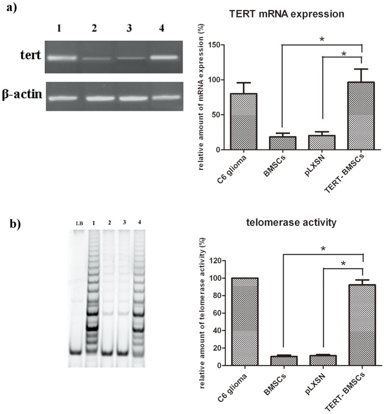
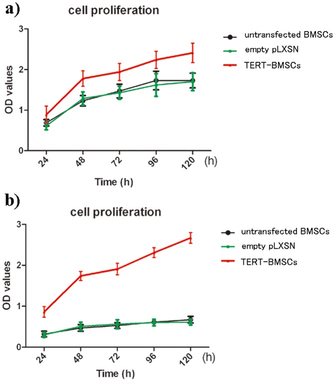
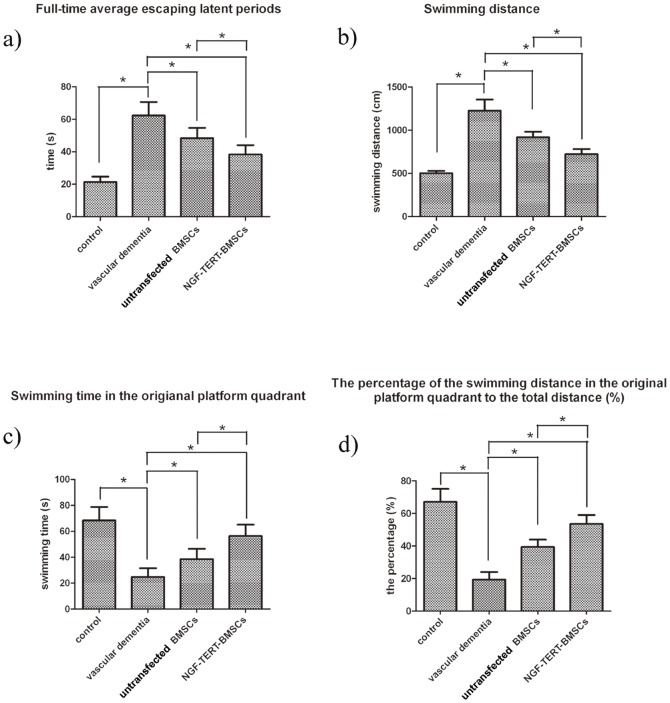
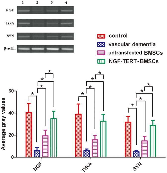
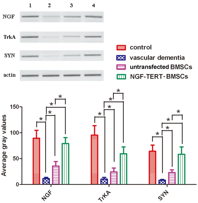
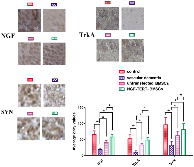
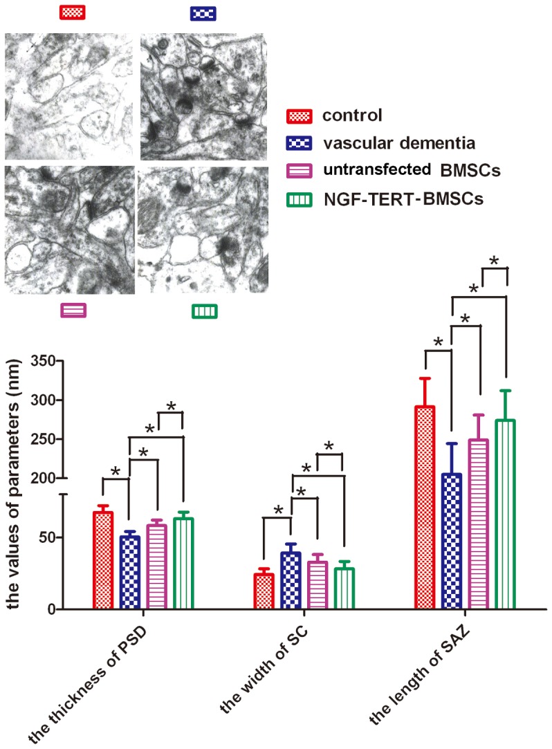
Vascular dementia (VaD) is a mental disorder caused by brain damage due to cerebrovascular disease, and incidence of VaD is rising. To date, there is no known effective cure for VaD, so effort in developing an effective treatment for VaD is of great importance. The differentiation plasticity of BMSCs, in conjunction with its weak immunogenicity, makes manipulated BMSCs an attractive strategy for disease treatment. However, BMSCs often display disabled differentiation, premature aging, and unstable proliferation, reducing their neuroprotective function. These problems may be caused by the lack of telomerase activity in BMSCs. Our results show that NGF-TERT co-transfected BMSCs have a better therapeutic effect than BMSCs lacking NGF and TERT expression, demonstrated by significant improvements in learning and memory in VaD rats. The underlying mechanism might be increased expression of NGF, TrkA and SYN in the hippocampal CA1 area, which has potential implication in advancing therapeutics for VaD.


Conflict of interest statement

Competing Interests: The authors have declared that no competing interests exist.

Figures

Figure 1
Figure 1. TERT mRNA expression and telomerase activity in TERT-BMSCs.
TERT mRNA expression and telomerase activity were significantly increased in TERT-BMSCs compared to untransfected BMSCs (P<0.05). a), tert and β-actin mRNA expression by RT-PCR analysis. b), telomerase activity analysis by TRAP. LB, lysis buffer (as a negative control); 1, C6 glioma cell line (as a positive control); 2, untransfected BMSCs; 3, BMSCs transfected with empty pLXSN; 4, TERT-BMSCs (transfected with recombinant TERT-pLXSN).
Figure 2
Figure 2. Cell proliferation analysis was performed after cell culture for 1 month (Figure 2a) and 2 months (Figure 2b).
The rate of proliferation in TERT-BMSCs was significantly increased compared to untransfected BMSCs and empty vector-transfected BMSCs following one month of cell culture (P<0.05) (Figure 2a). After long-term culture of 2 months, the rate of proliferation decreased in untransfected and empty-vector-transfected BMSCs but remained high in TERT-BMSCs (P<0.05) (Figure 2b).
Figure 3
Figure 3. The results of the Morris water maze test in four groups.
In the positioning navigation test, the full-time average escape latency period and swimming distance of untransfected and NGF-TERT co-transfected BMSC transplanted VaD groups during three consecutive days were significantly shorter than those of the VaD group (Figure 3a). Escape period and swimming distance of the NGF-TERT co-transfected BMSC transplanted VaD group were significantly shorter than those of the untransfected BMSC transplanted VaD group (Figure 3b). In the space exploration test, the VaD rats treated with untransfected or NGF-TERT co-transfected BMSCs showed significantly longer swimming time in the original platform quadrant and significantly higher percentages of swimming distance in the original platform quadrant than those of the VaD group (Figure 3c). Additionally, the VaD rats treated with NGF-TERT co-transfected BMSCs exhibited significantly longer swimming times and distances in the original quadrant than the rats treated with untransfected BMSCs (Figure 3d) (*P<0.05).
Figure 4
Figure 4. mRNA expression analysis by quantitative real-time RT-PCR.
mRNA expression levels of NGF/β-actin, TrkA/β-actin, and SYN/β-actin in the VaD group were significantly lower than those in the control group. Expression levels in untransfected and NGF-TERT co-transfected BMSC transplanted VaD groups were significantly higher than those of the VaD group. Additionally, expression levels in rats treated with NGF-TERT co-transfected BMSCs were significantly higher than those in rats treated with untransfected BMSCs (*, P<0.05, n = 12).
Figure 5
Figure 5. Protein expression analysis by western blotting.
After normalisation to actin, protein levels of NGF, TrkA, and SYN in the VaD group were significantly lower than those in the control group. The untransfected and NGF-TERT co-transfected BMSC transplanted VaD groups had significantly higher levels of these proteins than the VaD group. Additionally, rats treated with NGF-TERT co-transfected BMSCs had significantly higher levels of NGF, TrkA, and SYN than rats treated with untransfected BMSCs (*P<0.05, n = 12).
Figure 6
Figure 6. Immunohistochemical analysis of protein expression.
NGF-positive and TrkA-positive cells were detected in the hippocampus and cortex, and these proteins were most highly abundant in the cytoplasm of hippocampal cells. SYN protein expression was stronger in the emitting layer of the hippocampus CA1 area than in the molecular layer, and the outline of the cone cells showed minimal SYN. The average grey values of NGF, TrkA, and SYN proteins in the VaD group were significantly lower than those in the control group. The average grey values of untransfected and NGF-TERT co-transfected BMSCs transplanted VaD groups were higher than those of the VaD group. Additionally, grey values of NGF-TERT co-transfected BMSCs transplanted VaD rats were significantly higher than those of the untransfected BMSCs transplanted VaD rats (*P<0.05, n = 12).
Figure 7
Figure 7. Ultrastructure in the hippocampal CA1 area by transmission electron microscopy (×40,000).
The synaptic structural parameters of the hippocampal CA1 area in each group are shown. PSD, postsynaptic density; SC, synaptic cleft; SAZ, synaptic active zone. (*P<0.05, n = 12).

References

    1. Ikejima C, Yasuno F, Mizukami K, Sasaki M, Tanimukai S, et al. (2009) Prevalence and causes of early-onset dementia in Japan: a population-based study. Stroke 40: 2709–2714. - PubMed
    1. Iadecola C (2013) The pathobiology of vascular dementia. Neuron 80: 844–866. - PMC - PubMed
    1. Akinyemi RO, Mukaetova-Ladinska EB, Attems J, Ihara M, Kalaria RN (2013) Vascular risk factors and neurodegeneration in ageing related dementias: Alzheimer's disease and vascular dementia. Curr Alzheimer Res 10: 642–653. - PubMed
    1. Willyard C (2013) Stem cells: A time to heal. Nature 503: S4–6. - PubMed
    1. Li MD, Atkins H, Bubela T (2014) The global landscape of stem cell clinical trials. Regen Med 9: 27–39. - PubMed

Publication types