. 2014 May 12;5(3):65.
doi: 10.1186/scrt454.

Alterations of gene expression and protein synthesis in co-cultured adipose tissue-derived stem cells and squamous cell-carcinoma cells: consequences for clinical applications

Alterations of gene expression and protein synthesis in co-cultured adipose tissue-derived stem cells and squamous cell-carcinoma cells: consequences for clinical applications

Eva Koellensperger et al. Stem Cell Res Ther. .

Abstract

Introduction: This is the first study evaluating the interactions of human adipose tissue derived stem cells (ADSCs) and human squamous cell carcinoma cells (SCCs), with regard to a prospective cell-based skin regenerative therapy and a thereby unintended co-localization of ADSCs and SCCs.

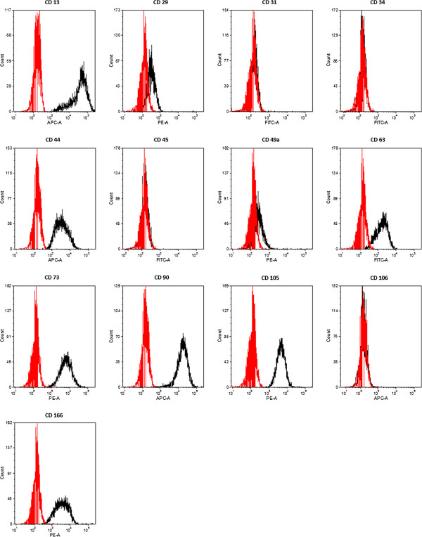
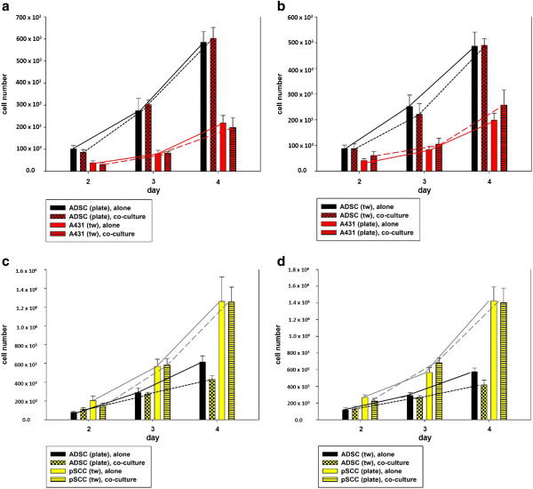
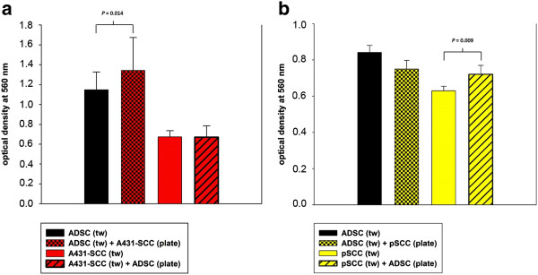
Methods: ADSCs were co-cultured with A431-SCCs and primary SCCs (pSCCs) in a transwell system, and cell-cell interactions were analyzed by assessing doubling time, migration and invasion, angiogenesis, quantitative real time PCR of 229 tumor associated genes, and multiplex protein assays of 20 chemokines and growth factors and eight matrix metalloproteinases (MMPS). Results of co-culture were compared to those of the respective mono-culture.

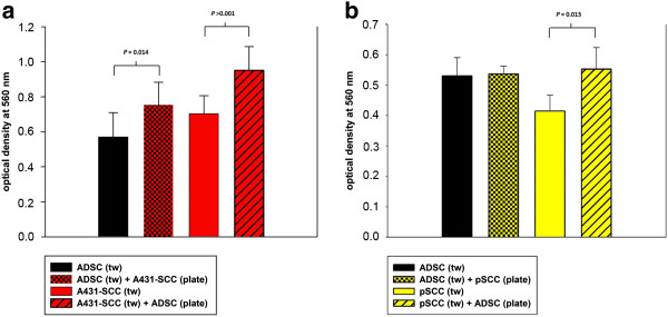
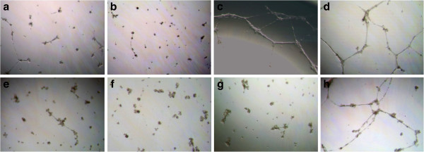
Results: ADSCs' proliferation on the plate was significantly increased when co-cultured with A431-SCCs (P = 0.038). PSCCs and ADSCs significantly decreased their proliferation in co-culture if cultured on the plate (P <0.001 and P = 0.03). The migration of pSCC was significantly increased in co-culture (P = 0.009), as well as that of ADSCs in A431-SCC-co-culture (P = 0.012). The invasive behavior of pSCCs and A431-SCCs was significantly increased in co-culture by a mean of 33% and 35%, respectively (P = 0.038 and P <0.001). Furthermore, conditioned media from co-cultured ADSC-A431-SCCs and co-cultured ADSCs-pSCCs induced tube formation in an angiogenesis assay in vitro.

Conclusions: This is the first study evaluating the possible interactions of primary human ADSCs with human SCCs, pointing towards a doubtlessly increased oncological risk, which should not be neglected when considering a clinical use of isolated human ADSCs in skin regenerative therapies.


Figures

Figure 1
Figure 1
Flow cytometry of pooled ADSCs from donors 1 to 6. Red lines show isotype controls, black lines show pooled ADSCs. ADSCs were positive for CD13, CD29, CD44, CD49a, CD63, CD73, CD90, CD105 and CD166. ADSCs were negative for CD31, CD34, CD45 and CD106. ADSCs, adipose tissue derived stem cells.
Figure 2
Figure 2
Representative light microscopical pictures of adipogenically and osteogenically differentiated ADSCs. Magnification 10x. (a) Intracellular lipid droplets stained by oil red method as a marker of adipogenic differentiation on Day 14 of differentiation. (b) Extracellular calcium deposition stained with alizarin red as a marker for osteogenic differentiation on Day 42 of differentiation. Undifferentiated controls are not shown. ADSCs, adipose tissue derived stem cells.
Figure 3
Figure 3
Effect of ADSC-SCC-co-culture on cells’ proliferative activity. (a) The growth of ADSCs on the plate significantly increased (P = 0.038) in co-culture with A431-SCCs in the transwell insert compared to mono-culture. The growth of co-cultured A431-SCCs in the transwell insert was not significantly affected (P >0.05). (b) ADSC-SCC-co-culture with A431-SCCs on the six-well plate and ADSCs in the transwell insert did not significantly affect cell growth of both cell types (P >0.05). (c) ADSC-SCC-co-culture with ADSCs on the six-well plate and pSCCs in the transwell insert significantly reduced ADSCs’ growth compared to mono-culture (P = 0.03). The proliferative activity of pSCCs in co-culture was not affected (P >0.05). (d) ADSC-SCC-co-culture with pSCCs on the six-well plate and ADSCs in the transwell insert significantly decreased the growth of pSCCs (P <0.001), that of co-cultured ADSCs was not significantly affected (P >0.05). ADSC, adipose tissue derived stem cells; SCCs, squamous cell carcinoma cells.
Figure 4
Figure 4
Migration assay. a) Migratory capacity of the ADSCs (black bar) and A431-SCCs (red bar) alone and in co-culture, measured as the level of optical density at 560 nm, with a standard deviation (SD). When co-cultured with A431-SCCs (black bar with red checkerboard pattern) ADSCs showed a significantly higher migration as in mono-culture (P = 0.014). Co-culture of A431-SCCs with ADSCs (red bars with black diagonal slashes) does not lead to a significant change in the migratory properties of A431-SCCs (P >0.05). b) Migratory capacity of the ADSCs (black bar) and pSCCs (yellow bar) alone and in co-culture, measured as the level of optical density at 560 nm, with a SD. When co-cultured with pSCCs (black bar with yellow checkerboard pattern) ADSCs showed a higher migration as in mono-culture (P >0.05). Co-culture of pSCCs with ADSCs (yellow bars with black diagonal slashes) resulted in a significantly increased migration of pSCCs (P = 0.009). ADSC, adipose tissue derived stem cells; pSCCs, primary squamous cell carcinoma cells; SCCs, squamous cell carcinoma cells.
Figure 5
Figure 5
Invasion assay. a) Invasive behavior of ADSCs (black bar) and A431-SCCs (red bar) alone and in co-culture, measured as the level of optical density at 560 nm with a standard deviation (SD). When co-cultured with A431-SCCs (black bar with red checkerboard pattern) ADSCs showed a significantly higher invasive capacity as in mono-culture (P = 0.014). Co-culture of SCCs with ADSCs (red bars with black diagonal slashes) also leads to a significant increase in the invasive behavior of A431-SCCs (P <0.001). b) Invasive behavior of ADSCs (black bar) and pSCCs (yellow bar) alone and in co-culture, measured as the level of optical density at 560 nm, with a SD. When co-cultured with pSCCs (black bar with yellow checkerboard pattern) ADSCs did not significantly change their invasive capacity compared to mono-culture. Co-culture of pSCCs with ADSCs (yellow bars with black diagonal slashes), however, leads to a significant increase in the invasive behavior of pSCCs (P = 0.013). ADSC, adipose tissue derived stem cells; pSCCs, primary squamous cell carcinoma cells; SCCs, squamous cell carcinoma cells.
Figure 6
Figure 6
Induction of angiogenesis. Angiogenesis assay with incubation of HUVEC cells with conditioned media from mono-cultured ADSCs (a, e), A431-SCCs (b), pSCCs (f), co-cultured ADSC-A431-SCCs (c), and co-cultured ADSCs-pSCCs (g). Controls (d, h) were induced with PMA. Tube formation could be detected with conditioned media from co-cultured ADSC-A431-SCCs, slightly from co-cultured ADSCs-pSCCs, and mono-cultured ADSCs or A431-SCCs. No significant angiogenesis could be detected when conditioned media from pSCC- or A431-SCC-mono-culture was added to the system. ADSC, adipose tissue derived stem cells; HUVEC, human umbilical vein endothelial cells; PMA, phorbol 12-myristate 13-acetate; pSCCs, primary squamous cell carcinoma cells; SCCs, squamous cell carcinoma cells.

Similar articles

Cited by

References

    1. Kim WS, Park BS, Park SH, Kim HK, Sung JH. Antiwrinkle effect of adipose-derived stem cell: activation of dermal fibroblast by secretory factors. J Dermatol Sci. 2009;53:96–102. doi: 10.1016/j.jdermsci.2008.08.007. - DOI - PubMed
    1. Kim WS, Park BS, Sung JH. The wound-healing and antioxidant effects of adipose-derived stem cells. Expert Opin Biol Ther. 2009;9:879–887. doi: 10.1517/14712590903039684. - DOI - PubMed
    1. Kim WS, Park BS, Sung JH. Protective role of adipose-derived stem cells and their soluble factors in photoaging. Arch Dermatol Res. 2009;301:329–336. doi: 10.1007/s00403-009-0951-9. - DOI - PubMed
    1. Kim WS, Park SH, Ahn SJ, Kim HK, Park JS, Lee GY, Kim KJ, Whang KK, Kang SH, Park BS, Sung JH. Whitening effect of adipose-derived stem cells: a critical role of TGF-beta 1. Biol Pharm Bull. 2008;31:606–610. doi: 10.1248/bpb.31.606. - DOI - PubMed
    1. Kim JH, Jung M, Kim HS, Kim YM, Choi EH. Adipose-derived stem cells as a new therapeutic modality for ageing skin. Exp Dermatol. 2011;20:383–387. doi: 10.1111/j.1600-0625.2010.01221.x. - DOI - PubMed

Publication types

MeSH terms