Review
. 2014 Nov;144(2):162-91.
doi: 10.1016/j.pharmthera.2014.05.012. Epub 2014 Jun 2.

The purinergic neurotransmitter revisited: a single substance or multiple players?

Affiliations
Review

The purinergic neurotransmitter revisited: a single substance or multiple players?

Violeta N Mutafova-Yambolieva et al. Pharmacol Ther. 2014 Nov.

Abstract

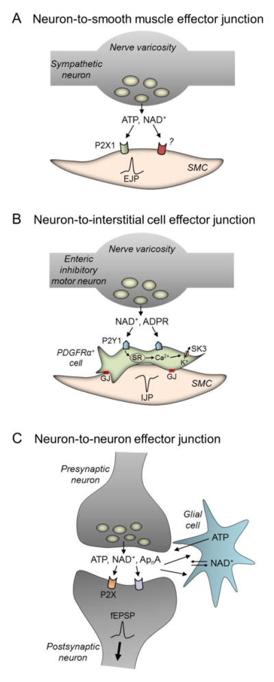
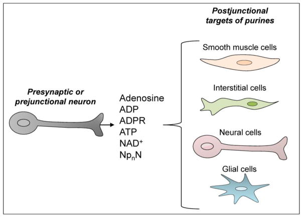
The past half century has witnessed tremendous advances in our understanding of extracellular purinergic signaling pathways. Purinergic neurotransmission, in particular, has emerged as a key contributor in the efficient control mechanisms in the nervous system. The identity of the purine neurotransmitter, however, remains controversial. Identifying it is difficult because purines are present in all cell types, have a large variety of cell sources, and are released via numerous pathways. Moreover, studies on purinergic neurotransmission have relied heavily on indirect measurements of integrated postjunctional responses that do not provide direct information for neurotransmitter identity. This paper discusses experimental support for adenosine 5'-triphosphate (ATP) as a neurotransmitter and recent evidence for possible contribution of other purines, in addition to or instead of ATP, in chemical neurotransmission in the peripheral, enteric and central nervous systems. Sites of release and action of purines in model systems such as vas deferens, blood vessels, urinary bladder and chromaffin cells are discussed. This is preceded by a brief discussion of studies demonstrating storage of purines in synaptic vesicles. We examine recent evidence for cell type targets (e.g., smooth muscle cells, interstitial cells, neurons and glia) for purine neurotransmitters in different systems. This is followed by brief discussion of mechanisms of terminating the action of purine neurotransmitters, including extracellular nucleotide hydrolysis and possible salvage and reuptake in the cell. The significance of direct neurotransmitter release measurements is highlighted. Possibilities for involvement of multiple purines (e.g., ATP, ADP, NAD(+), ADP-ribose, adenosine, and diadenosine polyphosphates) in neurotransmission are considered throughout.

Keywords: ADP-ribose; ATP; Adenosine; NAD; Nervous system; Purinergic neurotransmission.


Conflict of interest statement

Conflict of Interest Statement

The authors declare that there are no conflicts of interest.

Figures

Figure 1
Figure 1. Lack of fast excitatory junction potentials in guinea-pig mesenteric veins is not due to an absence of ATP release in response to nerve stimulation
A. Comparison of intracellular responses in [guinea-pig] mesenteric artery and vein to stimulation of lumbar colonic nerves. A1 and B1 are computer average of 25 consecutive responses evoked at 0.3 Hz. A2 and B2 are single traces of responses in same cells at a slower sampling rate. Stimulus artefacts in B2 are a result of variable capture of their peaks at slow sampling rates. Resting membrane potential in artery −69 mV, in vein −77 mV (reproduced from Kreulen, 1986, with permission from the American Physiological Society). B. Original chromatograms [obtained with HPLC-FLD analysis] illustrating the electrical field stimulation (EFS)-evoked overflow of adenine nucleotides and adenosine from superfused guinea-pig inferior mesenteric artery (a) and vein (b). Note that these chromatograms are made from samples collected from an artery (8.9 mg) and vein (2.2 mg) from the same animal. [When normalized to tissue weight EFS-evoked release of ATP in vein exceeded release of ATP in artery]. LU, luminescence units. Reproduced from Bobalova & Mutafova-Yambolieva, 2001a with permission from John Wiley and Sons Ltd.
Figure 2
Figure 2. Membrane depolarization-evoked release of NAD+, but not ATP, from NGF-differentiated rat PC12 cells requires intact SNAP-25-mediated exocytosis
A. Original chromatograms from samples collected during perfusion of the PC12 cells with either 5 mM KCl (pre-stimulation sample) or in the presence of 60 mM KCl for different time periods. KCl 60 mM evoked overflow of ATP, ADP, AMP, adenosine (ADO) and a mixture of β-NAD, cADPR, and ADPR. [β-NAD is the predominant compound in the β-NAD+cADPR+ADPR mixture as determined by HPLC fraction analysis: see original text in Yamboliev et al., 2009 for reference]. Inset: chromatographic peaks generated by etheno-derivatized purine standards (1 pmol each). Note that β-NAD has significantly lower fluorescence coefficient in generating eADPR than ATP, ADP, ADPR, AMP and ADO in generating corresponding e-purines. B. Excerpts of original chromatograms of superfusates from botulinum neurotoxin A (BoNT/A)-pretreated cells with an emphasis on ATP and the mixture of β-NAD, ADPR, and cADPR. C–H. Quantified data from 4–5 experiments expressed as the mean ± SEM. Data are averaged for each individual purine as well as for the sum of all purines (total purines). *P<0.05 vs. controls at 0′ (KCl 5 mM). One-way ANOVA followed by Bonferroni’s multiple comparison tests. #P<0.05 vs. 0′ in controls. Unpaired Student’s t-test. Note that the evoked release of most purines, except for ATP, was significantly reduced by BoNT/A. In BoNT/A-treated cells secretion of β-NAD+ADPR+cADPR at 5–20′ was below the initial levels. Reproduced from Yamboliev et al., 2009, with permission from John Wiley and Sons Ltd.
Figure 3
Figure 3. In human urinary bladder EFS evokes a prominent release of NAD+ that exceeds the release of ATP. Exogenous β-NAD inhibits the human detrusor muscle contractions
A. Electrical field stimulation (EFS; 0.1 ms, 15 V, 60 s) evokes frequency-dependent overflow of adenine nucleotides in human isolated bladder detrusor smooth muscle. Original chromatograms are shown from HPLC-FLD analysis of purines in tissue superfusate samples collected before (prestimulation, PS) and during EFS (ST) at 4 and 16 Hz. Aliquots were derivatized with 2-chloroacetaldehyde (80°C, pH 4, 40 min) to 1,N6-etheno (e)-nucleotides (i.e., eATP, eADP, eAMP) and 1,N6-etheno-(e)ADO. At all frequencies of stimulation, 1,N6-etheno-(e)ADPR with elution time of 11.2 min is also observed. eADPR stands for β-NAD, because β-NAD represents 90–95% of the eADPR peak as discussed in the main text of Breen et al., 2006. The EFS (16 Hz)-evoked release of purines is significantly reduced in the presence of TTX (0.3 μmol/l for 30 min). Scales apply to all chromatograms. Note that β-NAD has a significantly lower fluorescence coefficient in generating eADPR than ATP generating eATP as demonstrated in Figure 2 inset. B. Original traces showing that exogenous β-NAD (1 μmol/l) inhibits both the frequency and amplitude of muscle contractions in a human detrusor muscle segment exhibiting spontaneous contractile activity. Reproduced from Breen et al., 2006, with permission from The American Physiological Society.
Figure 4
Figure 4. Direct stimulation of enteric motor nerve cell bodies evoked differential release of NAD+ and ATP in monkey colon; NAD+ was released from enteric nerve terminals in an action potential- and Ca2+-dependent manner whereas ATP was released from nerve cell bodies independent of action potentials and Ca2+ entry
A. Purines released by stimulation of serotonergic 5-HT3 receptors in monkey colon whole muscle preparations. (A) Chromatograms of tissue superfusates collected before (control) and during activation of 5-HT3 receptors with SR57227 (500 μmol L−1 for 30 s) in the absence and presence of ondansetron (10 μmol L−1, 30 min), tetrodotoxin (TTX, 0.5 μmol L−1, 30-min superfusion), or ω-conotoxin GVIA (ω-CtxG, 50 nmol L−1, 30-min superfusion) in monkey colon. Small amounts of ATP, ADP, β-NAD+, AMP, and ADO were present in superfusate samples in the absence of agonist. Stimulation of 5-HT3 receptors with SR57227 evoked additional release of purines that was inhibited by the 5-HT3 receptor antagonist ondansetron. SR57227-evoked release of β-NAD+, but not ATP, was reduced by the neural blockers TTX and ω-CtxG. Scale applies to all chromatograms. LU, luminescence units. B. Averaged data are means ± SEM and show release of ATP, ADP, AMP, ADO, β-NAD+, and total purines (calculated as ATP+ADP+AMP+ADO+β-NAD+) during activation of 5-HT3 receptors with SR57227 (SR) and in the presence of ondansetron (Ond, 10 μmol L−1, 30 min), TTX (0.5L μmol L−1, 30 min), and ω-CtxG (50 nmol L−1, 30 min). Overflow (femtomoles per milligram of tissue) is the overflow during 5-HT3 receptor activation less spontaneous overflow. Each peak was calibrated to individual etheno-derivatized purine standards. Asterisks denote significant differences from SR57227-evoked release (i.e. control release) (*P < 0.05, **P < 0.01, ***P<.001); number of experiments in parenthesis. Note that the release of β-NAD+, but not ATP, was significantly inhibited by TTX and ω-CtxG. Reproduced from Durnin et al., 2013, with permission from John Wiley and Sons Ltd.
Figure 5
Figure 5. Schematic models of purinergic neurotransmission at three potential types of neuroeffector junctions
A. Neuron-to-smooth muscle effector junction. Purines (i.e., ATP) released from sympathetic nerve terminals (e.g., in vas deferens and blood vessels) and possibly from parasympathetic nerve terminals in the urinary bladder, act through P2X, mainly P2X1, receptors on smooth muscle cells (SMC) to mediate membrane depolarization (excitatory junction potentials, EJP) and contraction. In blood vessels P2X1 receptors are clustered in regions adjacent sympathetic nerve varicosities (Hansen et al., 1999; Vial & Evans, 2005), suggesting that released purines bind receptors in a limited volume in the interstitium, in a synapse-like space. In the vas deferens, however, P2X1 receptors do not appear to form clusters on SMC (Liang et al., 2001), and therefore, volume transmission might apply to this smooth muscle. There is evidence that NAD+ is released during stimulation of sympathetic nerves in blood vessels (Smyth et al., 2004) and from unidentified nerves in the urinary bladder (Breen et al., 2006). It is not clear if ATP and NAD+ are stored in and released from the same population of vesicles in whole tissues. Co-storage of both purines was demonstrated in vesicles from NGF-differentiated PC12 cells (Yamboliev et al., 2009). Differential modulation of ATP and NAD+ release in blood vessels by neuronal Ca2+ channel inhibitors has been shown as well (Smyth et al., 2009). The postjunctional receptor targets of NAD+ in blood vessels and bladder have not yet been determined. B. Neuron-to-interstitial cell effector junction. Purines can elicit muscle responses by first activating receptors localized on interstitial cells that are in close apposition to nerve varicosities and connect to SMC via gap junctions (GJ). There are at least two types of interstitial cells in the enteric nervous system: interstitial cells of Cajal and platelet-derived growth factor receptor alpha-positive (PDGFRα+) cells. Depicted here is purinergic inhibitory motor transmission in the enteric nervous system. NAD+ released from enteric inhibitory motor nerve terminals activates P2Y1 receptors localized on closely apposed PDGFRα+ cells. P2Y1 receptors mediate activation of phospholipase C and release of Ca2+ from intracellular stores (sarcoplasmic reticulum, SR) which activates small-conductance K+ channels (SK3). Inhibitory junction potentials (IJP) conduct to SMCs via GJ between PDGFRα+ cells and SMC. IJPs reduce excitability and cause muscle relaxation. ADPR also activates enteric P2Y1 receptors causing hyperpolarization and muscle relaxation. ADPR can be produced from NAD+ (Durnin et al., 2012b), but it is not known if ADPR is also released from enteric nerve varicosities. PDGFRα+ cells may also mediate purinergic relaxation in the urinary bladder via activation of P2Y1 receptors and SK channels (Lee et al., 2014). It is currently unknown whether the inhibitory effects of NAD+ in the bladder (Breen et al., 2006) are mediated by P2Y1 receptors and PDGFRα+ cells. Clustering of P2Y1 receptors on PDGFRα+ cells close to neurotransmitter release sites (Kurahashi et al., 2011) allows purinergic transmission to occur in a synapse-like space. C. Neuron-to-neuron effector junction (synapse). Neuron-to-neuron communication involves a direct action of released transmitters on receptors localized in the postsynaptic nerve membrane. In the central nervous system, ATP, NAD+, and diadenosine polyphosphates (ApnA) released from nerves could mediate fast synaptic transmission (fast excitatory postsynaptic potentials, fEPSP) by targeting P2X receptors localized on adjacent neural cells. Similarly, in the enteric nervous system purines mediate communication between interneurons via P2X receptors, predominantly P2X2 and P2X3 subtypes, on the adjacent nerve cells. Neuronal release of adenosine (not depicted) (Brundege & Dunwiddie, 1998; Manzoni et al., 1994; Lovatt et al., 2012) may mediate a widespread purinergic presynaptic inhibition. As depicted, purines could also target glial cells. Furthermore, glial cells release purines, including ATP and NAD+, which could target P2 receptors on nearby nerve cells. Studies have also suggested transport of NAD+ into glial cells (Verderio et al., 2001; Ying et al., 2003).
Figure 6
Figure 6. Multiple purine neurotransmitters act on various types of effector cells
Schematic model demonstrating that multiple purines can be released from neurons, which can act at different postjunctional target cell types, including smooth muscle cells, interstitial cells (e.g., interstitial cells of Cajal and platelet-derived growth factor receptor α-positive cells), other neural cells, and glial cells. The identity of neurotransmitter substance and target cells could vary between systems and species. ADP, adenosine 5′-diphosphate; ADPR, ADP-ribose; ATP, adenosine 5′-triphosphate; NAD+, nicotinamide adenine dinucleotide; NpnN, dinucleoside polyphosphate.

Similar articles

Cited by

References

    1. Abbracchio MP, Burnstock G, Boeynaems JM, Barnard EA, Boyer JL, Kennedy C, et al. International Union of Pharmacology LVIII: update on the P2Y G protein-coupled nucleotide receptors: from molecular mechanisms and pathophysiology to therapy. Pharmacol Rev. 2006;58:281–341. - PMC - PubMed
    1. Abbracchio MP, Burnstock G, Verkhratsky A, Zimmermann H. Purinergic signalling in the nervous system: an overview. Trends Neurosci. 2009;32:19–29. - PubMed
    1. Aberer W, Kostron H, Huber E, Winkler H. A characterization of the nucleotide uptake of chromaffin granules of bovine adrenal medulla. Biochem J. 1978;172:353–360. - PMC - PubMed
    1. Alberti E, Mikkelsen HB, Wang XY, Diaz M, Larsen JO, Huizinga JD, et al. Pacemaker activity and inhibitory neurotransmission in the colon of Ws/Ws mutant rats. Am J Physiol Gastrointest Liver Physiol. 2007;292:G1499–G1510. - PubMed
    1. Allcorn RJ, Cunnane TC, Kirkpatrick K. Actions of alpha, beta-methylene ATP and 6-hydroxydopamine on sympathetic neurotransmission in the vas deferens of the guinea-pig, rat and mouse: support for cotransmission. Br J Pharmacol. 1986;89:647–659. - PMC - PubMed

Publication types

MeSH terms