Review
. 2014 Jun 1;28(11):1143-58.
doi: 10.1101/gad.242511.114.

Mammary stem cells and the differentiation hierarchy: current status and perspectives

Affiliations
Review

Mammary stem cells and the differentiation hierarchy: current status and perspectives

Jane E Visvader et al. Genes Dev. .

Abstract

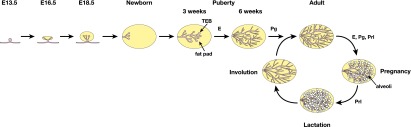
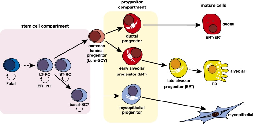
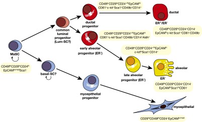
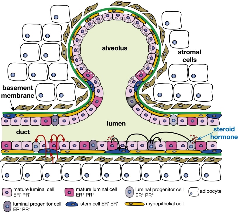
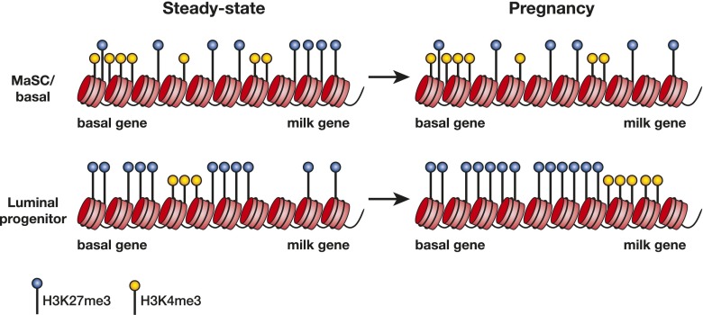
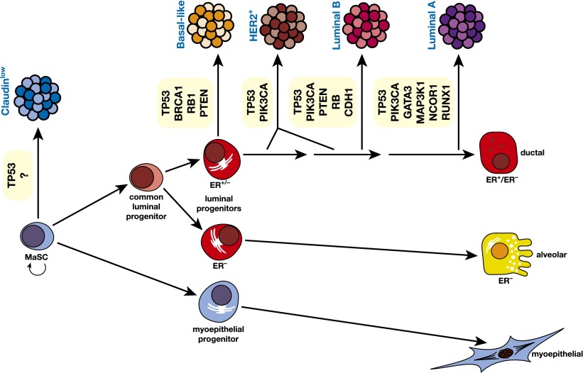
The mammary epithelium is highly responsive to local and systemic signals, which orchestrate morphogenesis of the ductal tree during puberty and pregnancy. Based on transplantation and lineage tracing studies, a hierarchy of stem and progenitor cells has been shown to exist among the mammary epithelium. Lineage tracing has highlighted the existence of bipotent mammary stem cells (MaSCs) in situ as well as long-lived unipotent cells that drive morphogenesis and homeostasis of the ductal tree. Moreover, there is accumulating evidence for a heterogeneous MaSC compartment comprising fetal MaSCs, slow-cycling cells, and both long-term and short-term repopulating cells. In parallel, diverse luminal progenitor subtypes have been identified in mouse and human mammary tissue. Elucidation of the normal cellular hierarchy is an important step toward understanding the "cells of origin" and molecular perturbations that drive breast cancer.

Keywords: breast cancer; development; epigenome; lineage tracing; mammary stem cell; steroid hormone.


Figures

Figure 1.
Figure 1.
Schematic diagram of the primary stages of mammary gland ontogeny in the embryo and adult. In the mouse embryo, the placodes (visible at embryonic day 11.5 [E11.5]) evolve into mammary buds that penetrate the underlying mesenchyme around E13.5. These buds sprout by E15.5 and develop a lumen. By E18.5, a small arborized gland that has invaded the developing fat pad is evident. In the postnatal animal, development of the mammary gland remains relatively dormant until puberty at 3 wk, when profound morphogenesis occurs, largely under the control of estrogen (E). In the young adult gland, progesterone (Pg) regulates side branching, while in pregnancy, the steroid hormones estrogen, progesterone, and prolactin (Prl) all play roles in alveolar expansion. In the late stages of pregnancy and during lactation, the peptide hormone prolactin plays a key role in establishing the secretory state. After lactation, the gland involutes and returns to a state that resembles the virgin gland.
Figure 2.
Figure 2.
Hypothetical model of the mammary epithelial hierarchy. A multipotent fetal MaSC has been identified. In the adult mammary gland, the stem cell compartment is heterogeneous and appears to comprise long-term and short-term repopulating cells (LT-RCs and ST-RCs, respectively), both of which are multipotent. These in turn give rise to committed progenitor cells for the myoepithelial and luminal (ductal and alveolar) epithelial lineages, but the precise number of progenitor cells is yet to be determined. Luminal progenitors are restricted to either a ductal or alveolar cell fate: The ductal progenitor possibly comprises both hormone receptor (HR)-positive and HR-negative cells, while the early and late alveolar-restricted progenitors are likely to be HR-negative. There may be a common luminal progenitor for these sublineages. The prospective isolation of cellular subsets from mouse and human mammary tissue provide support for the depicted hierarchical organization. In addition, two types of unipotent cells (lum-SC and myo-SC) may exist; current lineage tracing data are also consistent with long-lived progenitors performing these functions in vivo.
Figure 3.
Figure 3.
Markers of prospectively identified epithelial subsets in the mouse mammary gland. Summary of cell surface markers used for the isolation of epithelial cell subsets from the mouse mammary gland. ER denotes ERα.
Figure 4.
Figure 4.
Schematic depiction of potential regulatory cross-talk between different ductal mammary cells in response to steroid hormone stimulation. Estrogen or progesterone (red circles in lumen) activate ER+ epithelial cells (either mature cells or progenitors), which secrete paracrine factors that activate HR-negative stem cells or luminal progenitor cells and/or mature luminal cells. The black arrows indicate signals from steroid hormones, including those between HR-negative progenitors that have been indirectly activated by hormones, and their signaling to stem and other cells. The red arrows depict a further layer of interaction between stromal cells (fibroblasts and adipocytes) and mammary epithelial cells lining the ducts.
Figure 5.
Figure 5.
Regulation of the epigenetic state by steroid hormones and a dominant role for H3K27me3 modification. In the MaSC/basal population of the steady-state (virgin) gland, genes are epigenetically marked by H3K4me3 and H3K27me3, but as cells restrict to the luminal lineage, H3K27me3 modifications increase, suppressing gene expression. During pregnancy, where progesterone is a key hormone, only small changes occur in the H3K4me3-modified landscape of the MaSC/basal and luminal subsets, whereas a profound increase in H3K27me3 modifications occurs within the luminal subset, implying that H3K27me3 marks are important for regulating alveologenesis. Hypothetical histone modifications on putative basal and milk genes in the different subsets and hormonal states are shown.
Figure 6.
Figure 6.
Schematic model of the human breast epithelial hierarchy and potential relationships with breast tumor subtypes. The five major tumor types are shown linked to their closest normal epithelial counterpart based on gene expression profiling. The HER2+ subtype could originate through amplification of the HER2 locus in a luminal target cell that is either ER+ or ER. Examples of commonly mutated genes in the different subtypes of breast cancer are indicated.

References

    1. Anthony TE, Heintz N 2008. Genetic lineage tracing defines distinct neurogenic and gliogenic stages of ventral telencephalic radial glial development. Neural Dev 3: 30. - PMC - PubMed
    1. Asselin-Labat ML, Shackleton M, Stingl J, Vaillant F, Forrest NC, Eaves CJ, Visvader JE, Lindeman GJ 2006. Steroid hormone receptor status of mouse mammary stem cells. J Natl Cancer Inst 98: 1011–1014 - PubMed
    1. Asselin-Labat ML, Sutherland KD, Barker H, Thomas R, Shackleton M, Forrest NC, Hartley L, Robb L, Grosveld FG, van der Wees J, et al. 2007. Gata-3 is an essential regulator of mammary-gland morphogenesis and luminal-cell differentiation. Nat Cell Biol 9: 201–209 - PubMed
    1. Asselin-Labat ML, Vaillant F, Sheridan JM, Pal B, Wu D, Simpson ER, Yasuda H, Smyth GK, Martin TJ, Lindeman GJ, et al. 2010. Control of mammary stem cell function by steroid hormone signalling. Nature 465: 798–802 - PubMed
    1. Asselin-Labat ML, Sutherland KD, Vaillant F, Gyorki DE, Wu D, Holroyd S, Breslin K, Ward T, Shi W, Bath ML, et al. 2011. Gata-3 negatively regulates the tumor-initiating capacity of mammary luminal progenitor cells and targets the putative tumor suppressor caspase-14. Mol Cell Biol 31: 4609–4622 - PMC - PubMed

Publication types