. 2014 Aug;23(8):1102-12.
doi: 10.1002/pro.2496. Epub 2014 Jun 18.

Enzyme structure captures four cysteines aligned for disulfide relay

Affiliations

Enzyme structure captures four cysteines aligned for disulfide relay

Yair Gat et al. Protein Sci. 2014 Aug.

Abstract

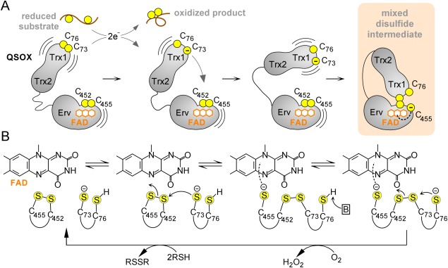
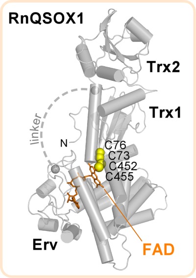
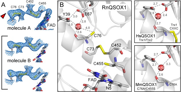
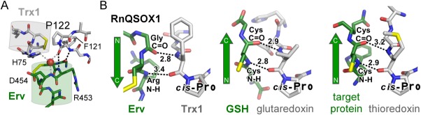
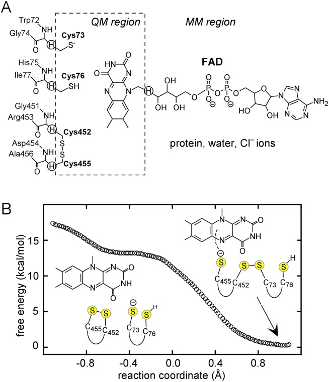
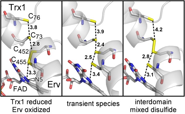
Thioredoxin superfamily proteins introduce disulfide bonds into substrates, catalyze the removal of disulfides, and operate in electron relays. These functions rely on one or more dithiol/disulfide exchange reactions. The flavoenzyme quiescin sulfhydryl oxidase (QSOX), a catalyst of disulfide bond formation with an interdomain electron transfer step in its catalytic cycle, provides a unique opportunity for exploring the structural environment of enzymatic dithiol/disulfide exchange. Wild-type Rattus norvegicus QSOX1 (RnQSOX1) was crystallized in a conformation that juxtaposes the two redox-active di-cysteine motifs in the enzyme, presenting the entire electron-transfer pathway and proton-transfer participants in their native configurations. As such a state cannot generally be enriched and stabilized for analysis, RnQSOX1 gives unprecedented insight into the functional group environments of the four cysteines involved in dithiol/disulfide exchange and provides the framework for analysis of the energetics of electron transfer in the presence of the bound flavin adenine dinucleotide cofactor. Hybrid quantum mechanics/molecular mechanics (QM/MM) free energy simulations based on the X-ray crystal structure suggest that formation of the interdomain disulfide intermediate is highly favorable and secures the flexible enzyme in a state from which further electron transfer via the flavin can occur.

Keywords: X-ray crystallography; cis-proline; enzyme mechanism; flavin adenine dinucleotide; quantum mechanics/molecular mechanics; thioredoxin fold.


Figures

Figure 1
Figure 1
QSOX catalytic cycle. (A) Schematic diagram of initial steps in QSOX catalysis. Juxtaposed yellow balls represent disulfide-bonded cysteines, and isolated balls represent reduced, unpaired cysteines. Structural domains of QSOX are labeled, and the FAD cofactor is shown as fused orange hexagons. (B) Active-site chemistry. Only the FAD cofactor and sulfur atoms of the CXXC motifs are shown explicitly. Cysteine protonation states at various steps are surmised, and the boxed “B” represents a generic base. An interactive view is available in the electronic version of the article.
Figure 2
Figure 2
Structure of RnQSOX1. Gray spheres mark the amino acids to either side of a flexible 12-residue segment for which electron density was not observed (“linker,” approximated as a dashed line). “N” is the protein amino terminus.
Figure 3
Figure 3
(A) Simulated annealing omit maps, contoured at 1 σ, in the vicinity of the RnQSOX1 CXXC motifs and FAD isoalloxazine. Cysteine residues and FAD are in stick representation. The alternate conformations that contribute to molecule B are bracketed. The red arrowhead indicates excess density in molecule A, which may indicate a minor population of partially reduced disulfides in this molecule as well. Electron density for the FAD isoalloxazine in molecule B is very poor, suggesting a mixture of redox states or conformational heterogeneity. (B) The putative proton transfer catalyst site of RnQSOX1, including Glu67, Tyr39, and a buried water molecule, is shown in relation to the cysteine ladder descending toward the FAD. Gray numbers are distances in Ångstrom. The N5 position of FAD is indicated by an arrowhead. Also shown is the coordination of the comparable buried water molecules in QSOX fragment and mutant structures (PDB codes 3Q6O and 3T38, respectively).
Figure 4
Figure 4
Interactions between the RnQSOX1 cis-proline and the target of nucleophilic attack. (A) The two RnQSOX1 CXXC motifs, at the amino-termini of their respective helices (cylinders), are shown in relation to cis-proline P122. Dashed lines indicate potential hydrogen bonds. Hydrogen bonds in black are shown in a different orientation in panel B. (B) Hydrogen bonding from the amino acid residue immediately upstream of the cis-proline to the substrate peptide is compared among RnQSOX1, a glutathione/glutaredoxin complex (PDB code 3FZ9), and a complex between thioredoxin and a target protein (PDB code 2IWT). Numbers indicate distances in Ångstrom.
Figure 5
Figure 5
(A) QM/MM partitioning scheme. Link atoms are marked by circled hydrogens. (B) Free energy profile for formation of the interdomain disulfide in RnQSOX1. The reaction coordinate (x-axis) is the antisymmetric stretch coordinate, defined as the difference between S–S bond distances: R(S452–S455)–R(S73–S452).
Figure 6
Figure 6
Snapshots from the free energy simulations, including the starting configuration, a transient species during formation of the interdomain disulfide, and the interdomain disulfide bridged state. Nonbonded sulfur–sulfur distances and distances between Cys455 and the FAD N5 position (arrowhead) are indicated in Ångstrom.

References

    1. Arnér ES, Holmgren A. Physiological functions of thioredoxin and thioredoxin reductase. Eur J Biochem. 2000;267:6102–6109. - PubMed
    1. Heras B, Shouldice SR, Totsika M, Scanlon MJ, Schembri MA, Martin JL. DSB proteins and bacterial pathogenicity. Nat Rev Microbiol. 2009;7:215–225. - PubMed
    1. Bulleid N, Ellgaard L. Multiple ways to make disulfides. Trends Biochem Sci. 2011;36:485–492. - PubMed
    1. Martin JL. Thioredoxin—a fold for all reasons. Structure. 1995;3:245–250. - PubMed
    1. Chivers PT, Prehoda KE, Raines RT. The CXXC motif: a rheostat in the active site. Biochemistry. 1997;36:4061–4066. - PubMed

Publication types