

Cervical spondylomyelopathy in Great Danes: a magnetic resonance imaging morphometric study
- PMID: 24888675
- PMCID: PMC4169205
- DOI: 10.1016/j.tvjl.2014.04.011
Cervical spondylomyelopathy in Great Danes: a magnetic resonance imaging morphometric study
Abstract
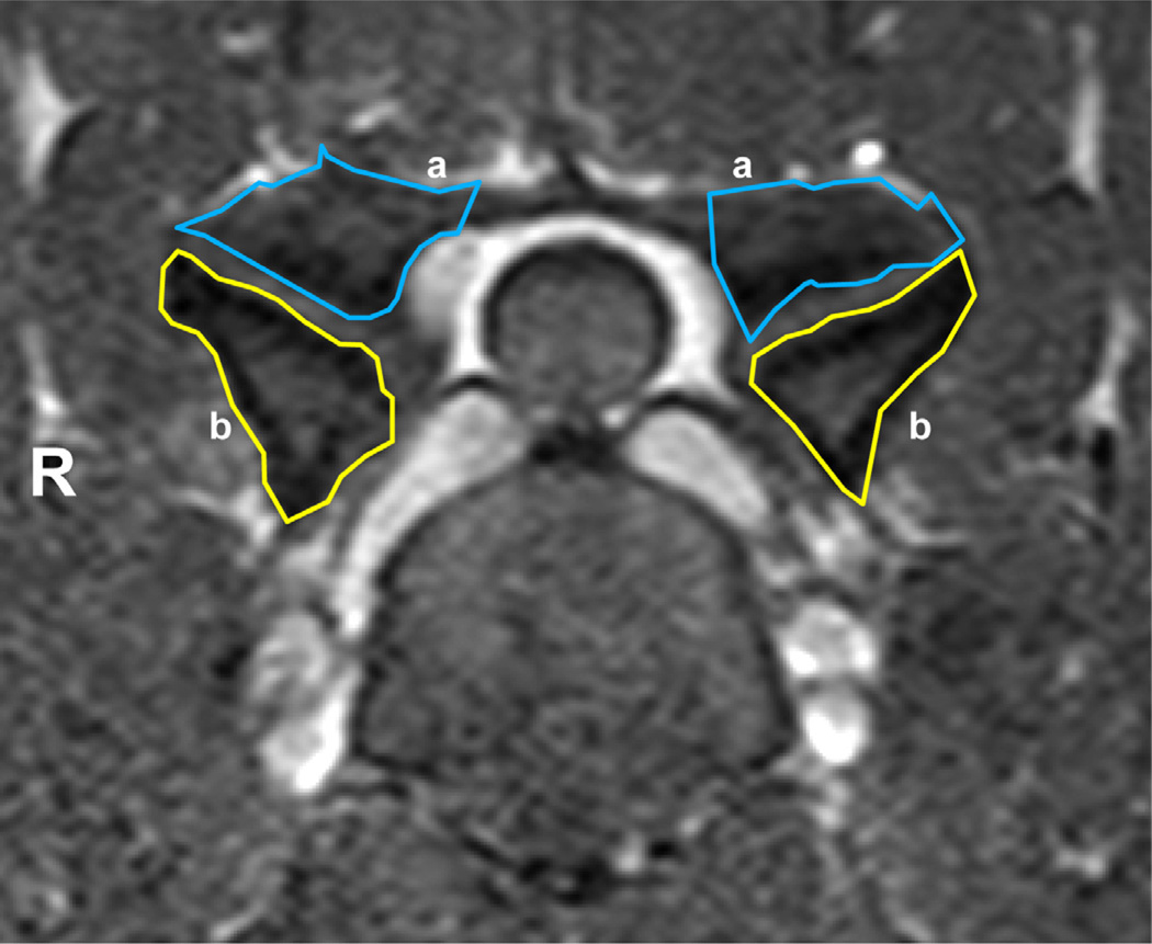
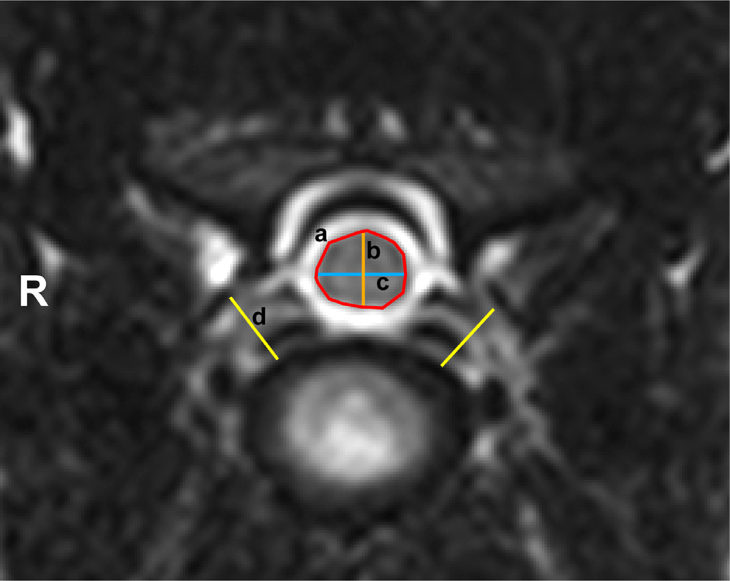
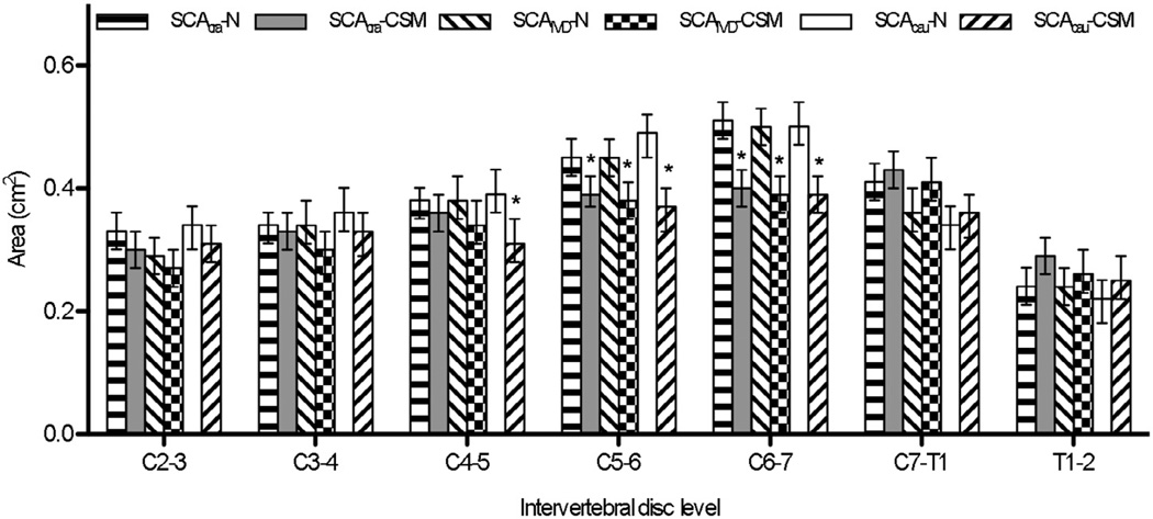
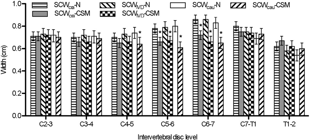
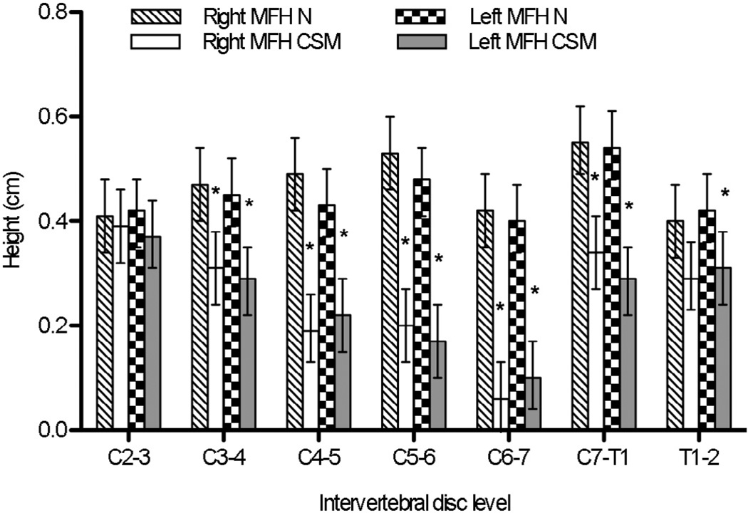
Morphometric investigations comparing normal and affected animals increase our understanding of spinal diseases in dogs. The aim of this study was to generate morphometric data for osseous-associated cervical spondylomyelopathy (CSM) in Great Danes (GDs). Magnetic resonance imaging (MRI) morphometric features of the cervical vertebral column of GDs with and without clinical signs of CSM were characterized and compared. Thirty client-owned GDs were prospectively enrolled, including 15 clinically normal and 15 CSM-affected GDs. All dogs underwent MRI of the cervical to thoracic vertebral column (C2-C3 through T1-T2). Areas of the cranial and caudal articular processes, and the height, width and areas of the vertebral canal and spinal cord were determined. Middle foraminal heights were measured. Intervertebral disc width was measured before and after traction. Intraobserver and interobserver agreement were calculated. CSM-affected GDs had larger areas of the caudal articular processes from C2-C3 through T1-T2. In CSM-affected GDs, the vertebral canal and spinal cord areas were significantly smaller at C5-C6 and C6-C7, the vertebral canal width was significantly narrower at C6-C7 and C7-T1, and the spinal cord width was significantly narrower at C5-C6 and C6-C7. Middle foraminal height was smaller in CSM-affected GDs from C3-C4 through C7-T1. Neutral intervertebral disc widths were smaller in CSM-affected GDs. It was concluded that the cervical vertebral canal dimensions are significantly different between normal and CSM-affected GDs. Absolute vertebral canal stenosis and severe foraminal stenosis involving the cervical vertebrae distinguish CSM-affected from clinically normal GDs. These findings are relevant to the pathogenesis of osseous-associated CSM and should be taken into consideration when performing imaging studies and planning surgery.
Keywords: Canine; Osseous-associated cervical spondylomyelopathy; Spinal cord; Stenosis; Wobbler syndrome.
Copyright © 2014 Elsevier Ltd. All rights reserved.
Conflict of interest statement
None of the authors has any financial or personal relationship that could inappropriately influence or bias the content of the paper.
Figures


Similar articles
-
Morphologic and morphometric magnetic resonance imaging features of Doberman Pinschers with and without clinical signs of cervical spondylomyelopathy.Am J Vet Res. 2006 Sep;67(9):1601-12. doi: 10.2460/ajvr.67.9.1601. Am J Vet Res. 2006. PMID: 16948609
-
Magnetic resonance imaging features of Great Danes with and without clinical signs of cervical spondylomyelopathy.J Am Vet Med Assoc. 2014 Aug 15;245(4):393-400. doi: 10.2460/javma.245.4.393. J Am Vet Med Assoc. 2014. PMID: 25075822 Free PMC article.
-
Comparison of angle, shape, and position of articular processes in Dobermans and Great Danes with and without cervical spondylomyelopathy.BMC Vet Res. 2017 Mar 24;13(1):77. doi: 10.1186/s12917-017-0997-4. BMC Vet Res. 2017. PMID: 28340590 Free PMC article.
-
Current insights and controversies in the pathogenesis and diagnosis of disc-associated cervical spondylomyelopathy in dogs.Vet Rec. 2012 Nov 24;171(21):531-7. doi: 10.1136/vr.e7952. Vet Rec. 2012. PMID: 23180710 Review.
-
Cervical spondylomyelopathy (wobbler syndrome) in dogs.Vet Clin North Am Small Anim Pract. 2010 Sep;40(5):881-913. doi: 10.1016/j.cvsm.2010.06.003. Vet Clin North Am Small Anim Pract. 2010. PMID: 20732597 Review.
Cited by
-
Clinical and magnetic resonance imaging characterization of cervical spondylomyelopathy in juvenile dogs.J Vet Intern Med. 2019 Sep;33(5):2160-2166. doi: 10.1111/jvim.15602. Epub 2019 Aug 30. J Vet Intern Med. 2019. PMID: 31469206 Free PMC article.
-
Thoracic vertebral canal stenosis in cats: clinical features, diagnostic imaging findings, treatment and outcome.J Feline Med Surg. 2020 Dec;22(12):1191-1199. doi: 10.1177/1098612X20920041. Epub 2020 May 21. J Feline Med Surg. 2020. PMID: 32436803 Free PMC article.
-
Long-term clinical and magnetic resonance imaging follow-up of dogs with osseous-associated cervical spondylomyelopathy.J Vet Intern Med. 2020 Sep;34(5):2012-2020. doi: 10.1111/jvim.15866. Epub 2020 Aug 14. J Vet Intern Med. 2020. PMID: 32794615 Free PMC article.
-
Diagnostic Imaging in Intervertebral Disc Disease.Front Vet Sci. 2020 Oct 22;7:588338. doi: 10.3389/fvets.2020.588338. eCollection 2020. Front Vet Sci. 2020. PMID: 33195623 Free PMC article. Review.
-
Cervical Intervertebral Disk to Vertebral Body Ratios of Different Dog Breeds Based on Sagittal Magnetic Resonance Imaging.Front Vet Sci. 2018 Oct 5;5:248. doi: 10.3389/fvets.2018.00248. eCollection 2018. Front Vet Sci. 2018. PMID: 30345279 Free PMC article.
References
-
- Boden SD, McCowin PR, Davis DO, Dina TS, Mark AS, Wiesel S. Abnormal magnetic-resonance scans of the cervical spine in asymptomatic subjects. Journal of Bone and Joint Surgery. 1990;72:1178–1184. - PubMed
-
- Bogduk N. Degenerative joint disease of the spine. Radiologic Clinics of North America. 2012;50:613–628. - PubMed
-
- Breit S, Künzel W. Shape and orientation of articular facets of cervical vertebrae (C3–C7) in dogs denoting axial rotational ability: An osteological study. European Journal of Morphology. 2002;40:43–51. - PubMed
-
- da Costa RC. Cervical spondylomyelopathy (Wobbler syndrome) in dogs. Veterinary Clinics of North America: Small Animal Practice. 2010;40:881–913. - PubMed
Publication types
MeSH terms
Grants and funding
LinkOut - more resources
Full Text Sources
Other Literature Sources
Medical
Miscellaneous
