Review
. 2015 Jan;467(1):175-90.
doi: 10.1007/s00424-014-1536-9. Epub 2014 Jun 3.

Mechanotransduction in the muscle spindle

Affiliations
Review

Mechanotransduction in the muscle spindle

Guy S Bewick et al. Pflugers Arch. 2015 Jan.

Abstract

The focus of this review is on the principal sensory ending of the mammalian muscle spindle, known as the primary ending. The process of mechanosensory transduction in the primary ending is examined under five headings: (i) action potential responses to defined mechanical stimuli-representing the ending's input-output properties; (ii) the receptor potential-including the currents giving rise to it; (iii) sensory-terminal deformation-measurable changes in the shape of the primary-ending terminals correlated with intrafusal sarcomere length, and what may cause them; (iv) putative stretch-sensitive channels-pharmacological and immunocytochemical clues to their identity; and (v) synaptic-like vesicles-the physiology and pharmacology of an intrinsic glutamatergic system in the primary and other mechanosensory endings, with some thoughts on the possible role of the system. Thus, the review highlights spindle stretch-evoked output is the product of multi-ionic receptor currents plus complex and sophisticated regulatory gain controls, both positive and negative in nature, as befits its status as the most complex sensory organ after the special senses.


Figures

Fig. 1
Fig. 1
The structure of the primary ending and its enclosing capsules, as illustrated by a representative transverse section (a; cat tenuissimus, 1-μm-thick section, toluidine blue stain; Ia-br myelinated banches of the Ia parent axon, ic inner capsule, imf intrafusal muscle fibre, oc outer capsule, ps periaxial space, st sensory terminal, short arrow myelinated Ia axon, long arrow nuclei of intrafusal nuclear bag fibre; scale bar = 20 μm.) and by reconstruction (b, c) from serial transverse sections, including that in (a). b Stereopair of complete ending, with terminals in shades of blue/violet distributed, by repeated branching of the parent Ia afferent nerve fibre (Ia Ia parent axon with myelin in two shades of grey; Schwann cell nuclei in red), to the seven intrafusal muscle fibres present in this case (bif bundle of intrafusal muscle fibres). The horizontal bar indicates the position of the transverse EM section shown in (a). c Stereopair of one of the first-order branches of the Ia afferent, its two second-order branches each with a heminode (arrows) and its sensory terminals distributed to one of the intrafusal muscle fibres. Total length of reconstruction (b, c) is 365 μm
Fig. 2
Fig. 2
Examples of muscle-spindle primary endings responding to trapezoidal (a, c) and sinusoidal (b, d) stretches applied to the tendon of the muscle (peroneus tertius of cat). a, b The reproducibility of the responses when five separate presentations of the stimuli are given to the same primary ending. The responses are superimposed and each response is indicated by different coloured symbols. c, d The similarity of responses from five primary endings in four different preparations. The data used to construct the figure were obtained by the method given in [39] and are taken from their unpublished results. The responses are presented as plots of instantaneous frequency in which each symbol corresponds to a single action potential and is positioned according to the time the action potential was recorded (abscissa) and the reciprocal of the time since the previous action potential (ordinate)
Fig. 3
Fig. 3
The receptor potential of a spindle primary ending (top trace) recorded from the Ia afferent fibre in a TTX-poisoned muscle spindle, relative depolarisation upwards, in response to a trapezoidal stretch (lower trace; duration of trace, 1.5 s). The various phases of the response are described according to Hunt et al. [40], who identified the pdm and the later part of the prm as due to voltage-dependent K channels [40]
Fig. 4
Fig. 4
The fine structure of the sensory terminals of a spindle primary ending (a, b) and their deformation in response to maintained stretch (c). a Transverse section through an intrafusal muscle fibre (m label is located in one of the fibre’s myonuclei) with an enclosing sensory terminal (t). Note: (i) the basal lamina (bl) of the muscle fibre that is continuous over the outer surface of the sensory terminal and (ii) cells of the inner capsule (ic). Part of the sensory terminal (black rectangle) is enlarged below the main image to show the corrugated nature of its plasmalemma (t) compared with the smooth membranes of the adjacent ic cells. ef elastic fibres. b Longitudinal section through an intrafusal muscle fibre (m again label is located in the fibre’s myonuclei), showing the lentiform profiles of the sensory terminals (t) in this plane. npa nonmyelinated preterminal axon, ps periaxial space. c Outline tracing of the section shown in (b), together with similar sections through the same type of intrafusal fibre from two other spindles. Mean lengths of 50 sarcomeres on either side of the primary ending indicate that the spindles were fixed at increasing amounts of maintained tension from top to bottom (2.20-, 2.50- and 2.55-μm sarcomere lengths, respectively). Corresponding deformation of the terminal profiles was clearest in the increasing mean radii of the terminal/muscle fibre interfaces (5.2, 20.1 and 31.9 μm, respectively). Examples of representative terminal profiles are shown enlarged on the right, with the increased flattening of the terminal/muscle fibre interface on each fibre indicated by an arrow [8]
Fig. 5
Fig. 5
Evidence for amiloride-sensitive ENaC family members in spindle sensory terminals. a Confocal immunofluorescence images of labelling for α, β, γ and δ ENaC (red) localises to the sensory terminals, double-labelled with synaptophysin (green). Synaptophysin labels the synaptic-like vesicles in the primary sensory terminals. b Stretch-evoked firing is inhibited by amiloride in a dose-dependent manner, in the range of 1–1,000 μM. c Similar effects are seen with other amiloride analogues, except hexamethyleneamiloride (HMA) [71]
Fig. 6
Fig. 6
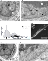
Fifty-nanometre, clear synaptic-like vesicle (SLV) clusters in spindle sensory terminals. a Electronmicrograph of a transverse section of the central portion of a nuclear bag intrafusal fibre (if) with its distinctive collection of prominent nuclei (n) and an enclosing sensory terminal (t). The boxed region is shown at higher magnification in (b), where distinctive clusters of synaptic-like vesicles can be seen (arrows), some aggregated towards and some away from, the muscle fibre. Quantification of vesicle diameters (c) shows the most abundant are clear and 50 nm (500 Å) in size, similar to their synaptic counterparts. Synapsin I labelling (d), a presynaptic vesicle-clustering protein, is present in the typical annulospiral ending of a rat lumbrical primary sensory terminal. Labelling in a motor nerve terminal in the same muscle is of similar intensity (inset, for comparison; NMJ, neuromuscular junction). Spindle terminals do not stain for synapsin II or III (Arild Njå, personal communication). Scale bar, 20 μm. e, f A coated pit of approximately 50-nm diameter in the axolemma of a sensory terminal, typical of endocytosis, as evidence of active SLV recycling. Note this pit is on the surface directed away from the nuclear bag fibre it encloses, although we have seen retrieval areas on both surfaces
Fig. 7
Fig. 7
FM1-43 labelling of differentiated primary spindle endings involves local synaptic-like vesicle recycling. Spontaneous FM1-43 labelling of primary endings in adult rat lumbrical muscle (a), showing characteristic differences in pitch, intrafusal fibre diameter and terminal ribbon width associated with nuclear bag (b) and chain (c) fibres. Incoming IA afferent axons also sequester dye (arrow) independent of activity due to their high myelin content. Intrafusal fibres enclosed by the endings are translucent, as they do not take up the dye. Terminal labelling is spontaneous but greatly increased by mechanical activity (repeated maximum stretch, b). It is also Ca2+ dependent, as it is essentially eliminated by the channel blocker Co2+ (c). d Unlike labelling by mechanosensory channel permeation, FM1-43 labelling in differentiated spindle terminals is reversible (d), showing clearly enhanced destaining with vibration (left images, 200 Hz, 50-μm amplitude, 5 min), a process which is also Ca2+ sensitive (right graph). This is consistent with FM1-43 uptake/release in differentiated terminals through local Ca2+-dependent recycling of SLVs in these endings ([16], b–d)
Fig. 8
Fig. 8
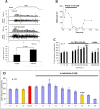
Endogenous glutamate secretion from SLVs maintains spindle stretch-evoked responsiveness. a A standard trapezoidal stretch-and-hold (~10 % muscle length) applied to a rat lumbrical muscle containing 8–12 muscle spindles evokes robust spiking activity in the electroneurogram (afferent discharge) and quantified in the firing frequency histogram (spike rate). Exogenous glutamate (1 h, 1 mM) can essentially double firing rate for the stretch. The histogram shows total firing within the 4-s plateau (hold phase) sample period indicated. Conversely, b inhibition of the highly atypical glutamate receptor with PCCG-13, applied in the absence of glutamate, can totally and reversibly block stretch-evoked spindle output. Note the timescale of hours, showing the long timecourse over which this modulation occurs. c Endogenous glutamate secretion occurs and is important for regulating firing, as blocking glutamate re-uptake by terminal excitatory amino acid transporters (TBOA), again in the absence of exogenous glutamate, enhances firing just as effectively as application of exogenous glutamate. *P < 0.05; ***P < 0.0001 vs. 30-min control firing (grey bars). 1- to 2-h wash reverses this effect (NS, not significantly different from pre-TBOA control). d Endogenous glutamate secretion is from SLVs. α-Latrotoxin, which evokes uncontrolled vesicle release, and ultimately vesicle depletion from spindle and synaptic endings [64], initially enhances stretch-evoked firing (*P < 0.05) then inhibits firing (***P < 0.0001), as SLVs are first released, then depleted. c1–c3 are recorded every 15 min, while t1–t10 are recorded at 30-min intervals. Btx-on bungarotoxin was first applied for 30 min prior to α-latrotoxin, to block spontaneous mechanical stimulation by fibre contraction driven by the α-latrotoxin-stimulated ACh secretion from fusimotor and extrafusal synaptic motor nerve terminals ([16], a, b)
Fig. 9
Fig. 9
Schematic summarising our current knowledge of the steps (1–7) from rest from mechanotransduction, through action potential encoding and firing rate determination, to autogenic sensitivity modulation. Areas of interest in each step are encircled or indicated by arrows.1, The myelinated primary afferent axon arrives from the left, produces a specialised encoding site at the unmyelinated heminode, then expands to form the sensory terminal proper, enclosing the intrafusal muscle fibre. The afferent discharge rate is shown in the panel bottom left (arrow). The terminal is the primary site of mechanotransduction through at least one type of mechanosensory channel (MS) passing Na+ and Ca2+. For convenience, these are shown separately (MSNC mechanosensitive Na+ channel, MSCC mechanosensitive Ca2+ channel). The terminal, as for all primary mechanosensory nerve endings, contains a population of 50-nm diameter clear vesicles—synaptic-like vesicles (SLVs, green circles—see text for details). At rest, SLVs undergo spontaneous exocytosis of glutamate (green dots in dotted area) to activate the phospholipase d-coupled metabotropic glutamate receptor (PLD-mGluR), to enable and maintain ending ability to respond to stretch stimuli. Abbreviations: Ca P/Q P/Q-type voltage-dependent Ca2+ channel, K Ca Ca2+-activated potassium channel, Na v voltage-dependent sodium channel. 2, Muscle stretch (green arrows) gates the MSNC, and Na+ influx depolarises the terminal. 3, The depolarisation spreads electrotonically to the much narrower heminode encoding region, increasing action potential (AP) firing (black arrow). Only the voltage-dependent Na+ channel component of the AP is shown for simplicity. 4, The APs trigger the opening of P/Q-type Ca2+ channels. 5, The resulting Ca2+ influx opens Ca2+-activated K+ channels (K Ca), repolarising the heminode region. This negative feedback step moderates the firing rate (black arrow). 6, Simultaneously, the initial stretch also gates a mechanosensitive Ca2+ current (through the MSNC or another mechanosensory channel (MSCC)), allowing Ca2+ influx. 7, The increased intracellular Ca2+ enhances SLV exocytosis of glutamate, further activating the PLD-mGluRs. The resulting increase in PLD activity (black arrow) is part of a positive feedback loop (curved arrows) that maintains the ability of the ending to respond to subsequent stretches, perhaps by enhancing/maintaining MS channel insertion, through a mechanism that awaits identification. An animated version of this sequence is available online (see Supplementary material, S1)
Fig. 10
Fig. 10
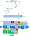
ad Progressive geometrical abstraction of a single terminal of a spindle primary ending, leading to a flow-chart summarising the events of mechanosensory transduction. Green block arrows in (ac) indicate the direction and distribution of stretch applied to the terminal when the primary ending is lengthened during muscle stretch or fusimotor stimulation. a A single terminal in its annulospiral form, taken from a primary ending reconstructed from serial sections [8]. Several such terminals typically enclose a single intrafusal muscle fibre. The terminal is connected to its associated heminode by a short, unmyelinated preterminal axonal branch at the point shown. b The terminal unrolled and turned through 90°. Note that individual terminals may be repeatedly branched and that the direction of stress during stretch is orthogonal to the long axis of the terminal. c A terminal and its associated unmyelinated preterminal branch shown in abstract cylindrical form to indicate the relative diameters of these structures. The smaller preterminal branch to the right is about 1 μm diameter. The lengths, especially that of the much larger terminal to the left, are highly variable. d Flow chart to illustrate the main events of mechanosensory transduction, as described in this review. The principal feed-forward pathway from stimulus (stretch) to output (action potentials) is shown by the white block arrows. We envisage that the overall gain of this pathway is controlled by several feedback pathways: negative feedback 1 is at present hypothetical and is included to account for the reversible silencing of the primary ending by PCCG-13 inhibition of the PLD-linked mGluR; the positive feedback pathway is the well-established SLV/glutamatergic loop; negative feedbacks 2 and 3 involve different kinds of K[Ca], one located in the terminal, the other in the heminode and both perhaps triggered by action potentials opening voltage-gated Ca channels. Green lines and arrowheads indicate enhancing/excitatory actions; red lines and circles indicate reducing/inhibitory actions

References

    1. Adrian ED, Zotterman Y. The impulses produced by sensory nerve endings: part II. The response of a single end-organ. J Physiol. 1926;61:151–171. doi: 10.1113/jphysiol.1926.sp002281. - DOI - PMC - PubMed
    1. Aguado F, Majó G, Ruiz-Montasell B, Llorens J, Marsal J, Blasi J. Syntaxin 1A and 1B display distinct distribution patterns in the rat peripheral nervous system. Neuroscience. 1999;88:437–446. doi: 10.1016/S0306-4522(98)00247-4. - DOI - PubMed
    1. Akoev GN, Alekseev NP, Krylov BV. Mechanoreceptors: their functional organisation. Berlin: Springer-Verlag; 1988.
    1. Althaus M, Bogdan R, Clauss WG, Fronius M. Mechano-sensitivity of epithelial sodium channels (ENaCs): laminar shear stress increases ion channel open probability. FASEB J. 2007;21:2389–2399. doi: 10.1096/fj.06-7694com. - DOI - PubMed
    1. Árnadóttir J, O'Hagan R, Chen Y, Goodman MB, Chalfie M. The DEG/ENaC protein MEC-10 regulates the transduction channel complex in Caenorhabditis elegans touch receptor neurons. J Neurosci. 2011;31:12695–12704. doi: 10.1523/JNEUROSCI.4580-10.2011. - DOI - PMC - PubMed

LinkOut - more resources